Copyright
©The Author(s) 2005.
World J Gastroenterol. Aug 14, 2005; 11(30): 4618-4622
Published online Aug 14, 2005. doi: 10.3748/wjg.v11.i30.4618
Published online Aug 14, 2005. doi: 10.3748/wjg.v11.i30.4618
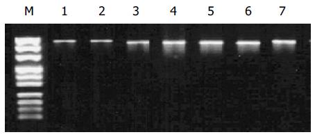
Figure 1 RT-PCR amplification of polβ gene.
Lanes 1, 2 and 7: Normal size of PCR products of H1, H2 and H3; lanes 3-6: shorter size of PCR product of H5, H8, H10, and H16; M: DNA marker (from top to bottom: 1 000, 900, 800, 700, 600, 500, 400, 300, 200, 100 bp).
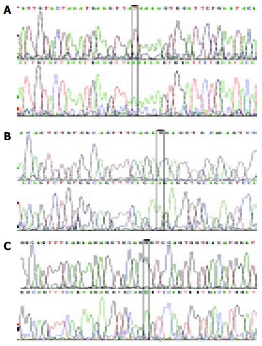
Figure 2 Mutations in the polβ gene.
A: 660 nt A→G mutation in H4 carcinoma; B: 670 nt A→G mutation in N1 carcinoma; C: 613 nt A→T mutation in H28 carcinoma.
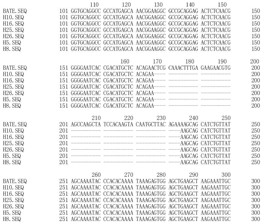
Figure 3 Comparison between wild type polβ gene fragment and six gene fragments with deletion (177→234 nt).
- Citation: Zhao GQ, Wang T, Zhao Q, Yang HY, Tan XH, Dong ZM. Mutation of DNA polymerase β in esophageal carcinoma of different regions. World J Gastroenterol 2005; 11(30): 4618-4622
- URL: https://www.wjgnet.com/1007-9327/full/v11/i30/4618.htm
- DOI: https://dx.doi.org/10.3748/wjg.v11.i30.4618


