Copyright
©The Author(s) 2005.
World J Gastroenterol. Jul 21, 2005; 11(27): 4287-4289
Published online Jul 21, 2005. doi: 10.3748/wjg.v11.i27.4287
Published online Jul 21, 2005. doi: 10.3748/wjg.v11.i27.4287
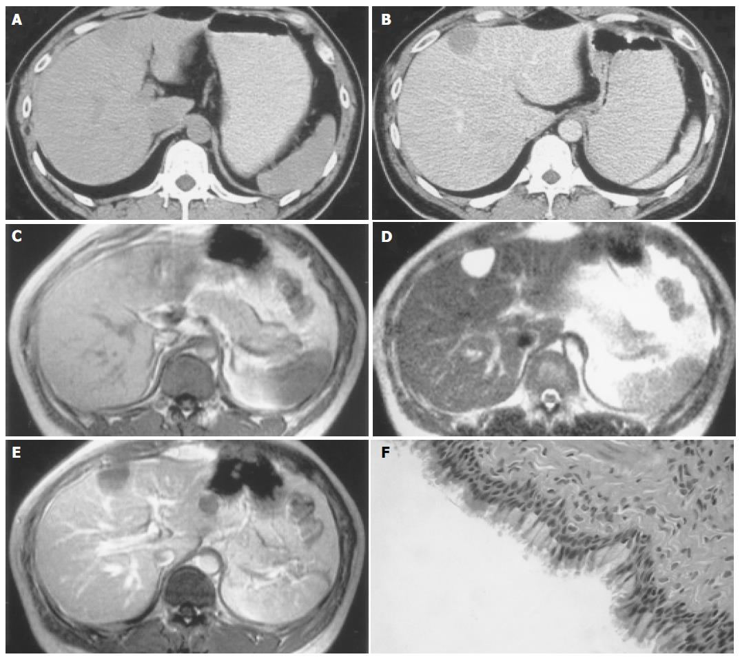
Figure 1 CHFC in a 30-year-old man.
A: Unenhanced CT scan shows a slightly hypoattenuating (47 HU) mass in the medial segment beneath the hepatic surface; B: On enhanced CT scan, a well-defined hypoattenuating mass is revealed with no enhancement; C: On axial T1-weighted imaging, the lesion appears isointense relative to surrounding liver parenchyma; D: Axial T2-weighted imaging shows markedly homogeneously hyperintense mass in the left liver; E: Enhanced T1-weighted imaging delineates slightly hypointense mass just beneath the hepatic surface; F: Photomicrograph reveals a cyst lined by ciliated pseudostratified columnar epithelial cells (HE × 400).
- Citation: Fang SH, Dong DJ, Zhang SZ. Imaging features of ciliated hepatic foregut cyst. World J Gastroenterol 2005; 11(27): 4287-4289
- URL: https://www.wjgnet.com/1007-9327/full/v11/i27/4287.htm
- DOI: https://dx.doi.org/10.3748/wjg.v11.i27.4287


