Copyright
©The Author(s) 2005.
World J Gastroenterol. Jul 14, 2005; 11(26): 4111-4113
Published online Jul 14, 2005. doi: 10.3748/wjg.v11.i26.4111
Published online Jul 14, 2005. doi: 10.3748/wjg.v11.i26.4111
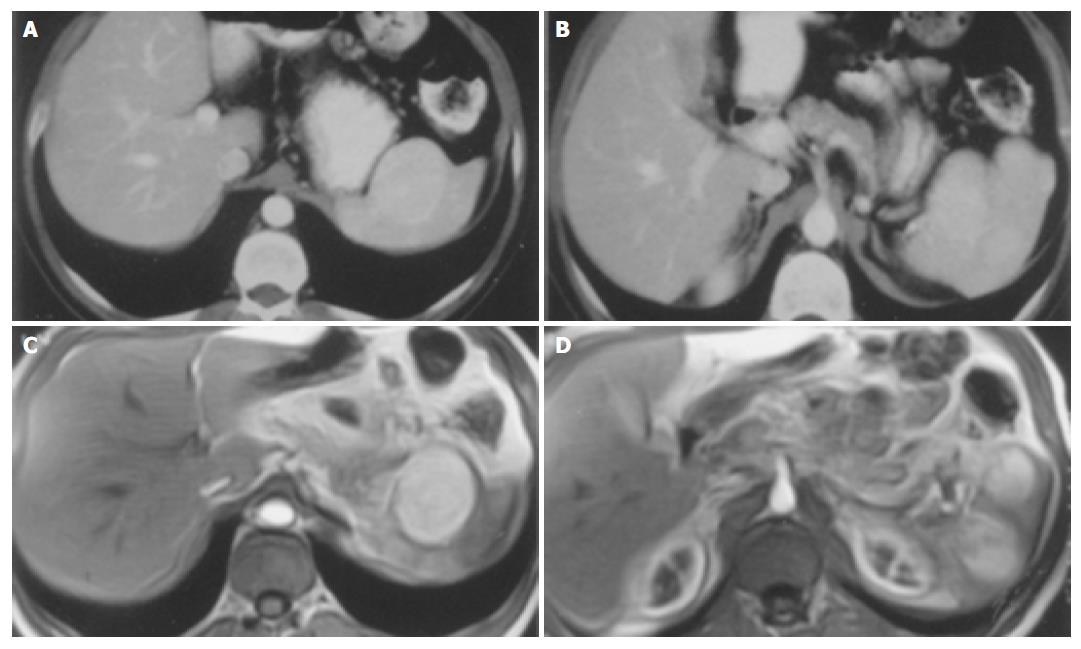
Figure 1 CT and MR images of splenic hemangiopericytoma.
A and B: Enhanced computed tomography scan showing high-density masses located in the spleen. C and D: T1-weighted abdominal magnetic resonance images showing multifocal hypervascular lesions in the spleen.
Figure 2 A: Spindle-shaped cells of hemangiopericytoma with eosinophilic cytoplasm around the capillary vessels in the spleen (HE ×40); B: (HE ×200); C: Dilated vessels of the cavernous hemangioma of the colonic serosa (HE × 40).
- Citation: Yilmazlar T, Kirdak T, Yerci O, Adim SB, Kanat O, Manavoglu O. Splenic hemangiopericytoma and serosal cavernous hemangiomatosis of the adjacent colon. World J Gastroenterol 2005; 11(26): 4111-4113
- URL: https://www.wjgnet.com/1007-9327/full/v11/i26/4111.htm
- DOI: https://dx.doi.org/10.3748/wjg.v11.i26.4111


