Copyright
©The Author(s) 2005.
World J Gastroenterol. Jul 14, 2005; 11(26): 4061-4066
Published online Jul 14, 2005. doi: 10.3748/wjg.v11.i26.4061
Published online Jul 14, 2005. doi: 10.3748/wjg.v11.i26.4061
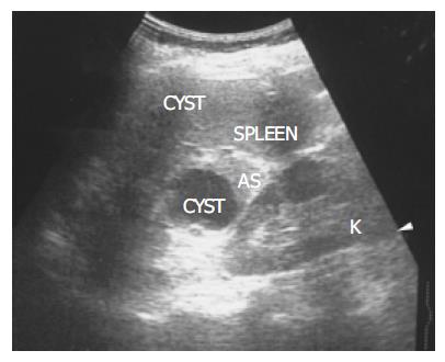
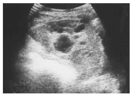
Figure 1 There is a homogenous, round contour near the hilum, identified as an accessory spleen (AS) in a 62-year-old female.
One anechoic cyst is noted within the accessory spleen. The pathologic process affecting the spleen (small cyst) also affects the accessory spleen (large cyst).
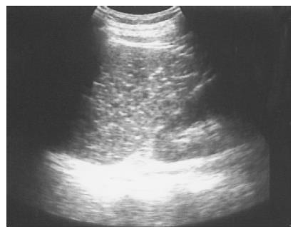
Figure 2 Multiple tiny calcified spots involving almost the entire spleen are found in a 58-year-old female with liver cirrhosis, called Gamna-Gandy bodies.
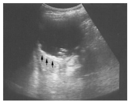
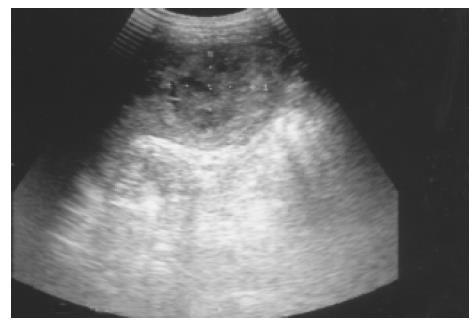
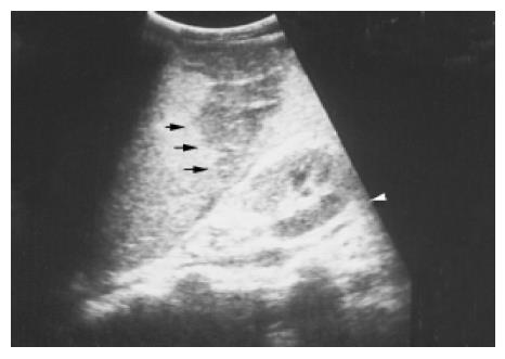
Figure 3 A pseudocyst cyst of the spleen after trauma reveals debris or echogenic contents within the thick wall (arrows) in a 60-year-old female.
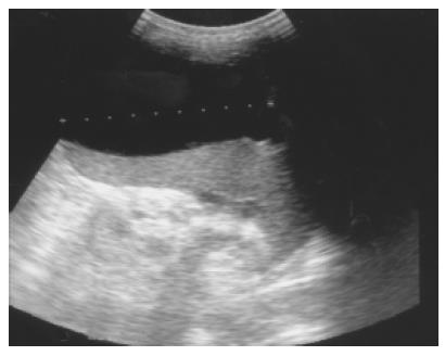
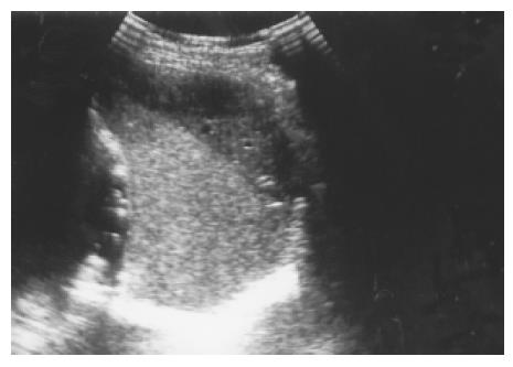
Figure 4 Pancreatic pseudocyst eroding into the adjacent spleen and mimicking a huge splenic simple cyst was noted after an episode of acute alcoholic pancreatitis in a 32-year-old male patient.
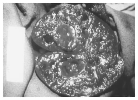
Figure 5 An atypical hemangioma can have mixed echogenicity with a dominant cystic portion.
Mild through transmission points to the cystic nature of the hemangioma.
Figure 6 Large and multiple hemangiomas occupy the entire spleen.
The hypoechoic areas shown in Figure 5 are filled with blood clots and thrombosis. The 48-year-old male received splenectomy due to LUQ pain caused by venous thrombosis in spleen.
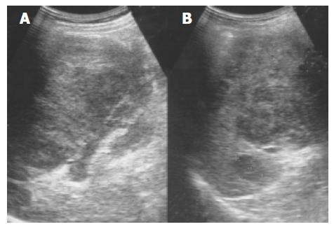
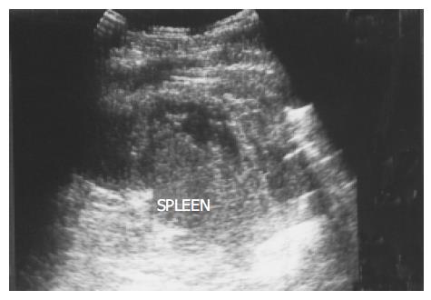
Figure 7 An enlarged spleen (A) demonstrates a diffusely coarse echotexture with several mixed echoic lesions (B) in a 58-year-old male patient.
Figure 8 Splenic metastasis from a lung carcinoma in a 68-year-old male is seen as a hypoechoic mass with a target sign.
Figure 9 A peripheral wedge-shaped hypoechoic region (arrows) was noted at the upper pole of spleen from an infarction after transcatheter arterial embolization in a 45-year-old male with hepatoma.
Figure 10 A subcapsular crescent-shaped hypoechoic lesion is noted at the upper pole of spleen.
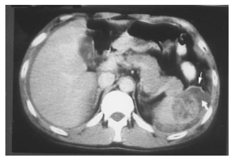
Figure 11 Several linear hypoechoic foci and a subcapsular fluid accumulation are noted in a spleen.
Loss of the normal architecture was seen after a traffic accident in a 38-year-old male.
Figure 12 A splenic laceration and rupture (in Figure 11) is identified as a blood-filled cleft (lower arrow) and capsular rupture (upper arrow) on contrast CT.
- Citation: Chen MJ, Huang MJ, Chang WH, Wang TE, Wang HY, Chu CH, Lin SC, Shih SC. Ultrasonography of splenic abnormalities. World J Gastroenterol 2005; 11(26): 4061-4066
- URL: https://www.wjgnet.com/1007-9327/full/v11/i26/4061.htm
- DOI: https://dx.doi.org/10.3748/wjg.v11.i26.4061


