Copyright
©The Author(s) 2005.
World J Gastroenterol. Jul 7, 2005; 11(25): 3866-3870
Published online Jul 7, 2005. doi: 10.3748/wjg.v11.i25.3866
Published online Jul 7, 2005. doi: 10.3748/wjg.v11.i25.3866
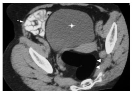
Figure 1 Optimal visualization of normal rectal wall (arrowheads), small intestine (arrow), and urinary bladder (star).
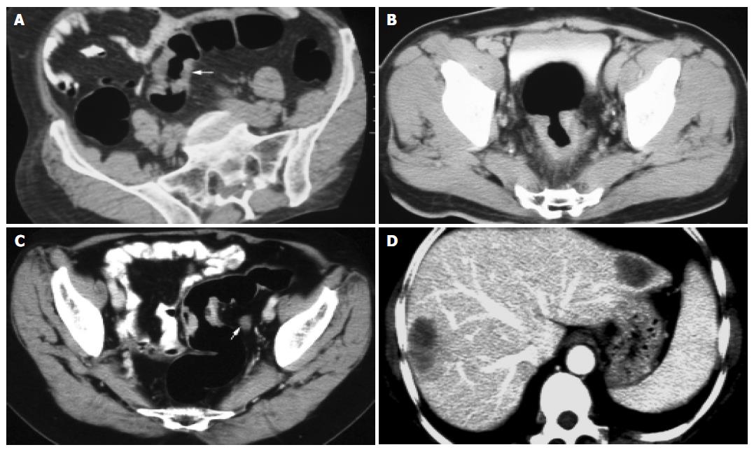
Figure 2 Correctly staged lesions.
A: Dukes stage A carcinoma (arrow); B: Dukes stage B carcinoma; C: Dukes stage C carcinoma; D: Dukes stage D carcinoma.
Figure 3 Incorrectly staged lesions.
A: Dukes stage A carcinoma; B: Dukes stage B carcinoma (arrow); C: Dukes stage B carcinoma.
- Citation: Sun CH, Li ZP, Meng QF, Yu SP, Xu DS. Assessment of spiral CT pneumocolon in preoperative colorectal carcinoma. World J Gastroenterol 2005; 11(25): 3866-3870
- URL: https://www.wjgnet.com/1007-9327/full/v11/i25/3866.htm
- DOI: https://dx.doi.org/10.3748/wjg.v11.i25.3866


