©2005 Baishideng Publishing Group Inc.
World J Gastroenterol. Jun 14, 2005; 11(22): 3331-3334
Published online Jun 14, 2005. doi: 10.3748/wjg.v11.i22.3331
Published online Jun 14, 2005. doi: 10.3748/wjg.v11.i22.3331
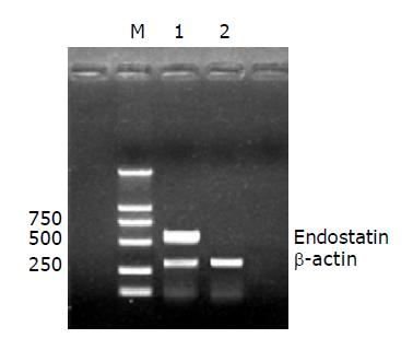
Figure 1 RT-PCR analysis of endostatin in transfected cells.
M: DL2000 DNA marker; lane 1: PCR products amplified from rAAV2-hEndo transfected cells; lane 2: PCR products amplified from AAV2 transfected cells.
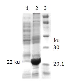
Figure 2 SDS-PAGE of the supernatant of transfected cells.
Lane 1: concentrated supernatant of AAV2 transfected Hep3B cells; lane 2: rAAV2-hEndo transfected Hep3B cells; lane 3: molecular weight standard (Amersham Biosciences), myosin (Mr 220000), phosphorylase (Mr 97000), albumin (Mr 66000), ovalbumin (Mr 45000), carbonic anhydrase (Mr 30000), trypsin inhibitor (Mr 20100), lysozyme (Mr 14300).
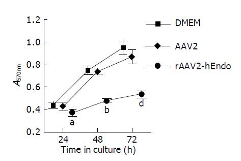
Figure 3 Proliferation inhibition of endothelial cells by supernatant from transfected and untransfected cells.
aP<0.05, bP<0.01 and dP<0.01 vs AAV2-transfected group.
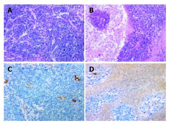
Figure 4 Immunohistochemical and HE staining of HCC xenograft.
A: AAV2-transfected tissue (HE staining, ×200); B: endostatin-transfected tissue (HE staining, ×200); C: MVD of AAV2-transfected tissue (stained with factor VIII-related antigen, ×200); D: MVD of rAAV2-hEndo transfected tissue (stained with factor VIII-related antigen, ×200).
- Citation: Liu H, Peng CH, Liu YB, Wu YL, Zhao ZM, Wang Y, Han BS. Inhibitory effect of adeno-associated virus-mediated gene transfer of human endostatin on hepatocellular carcinoma. World J Gastroenterol 2005; 11(22): 3331-3334
- URL: https://www.wjgnet.com/1007-9327/full/v11/i22/3331.htm
- DOI: https://dx.doi.org/10.3748/wjg.v11.i22.3331


