Copyright
©2005 Baishideng Publishing Group Inc.
World J Gastroenterol. Jun 7, 2005; 11(21): 3329-3329
Published online Jun 7, 2005. doi: 10.3748/wjg.v11.i21.3329
Published online Jun 7, 2005. doi: 10.3748/wjg.v11.i21.3329
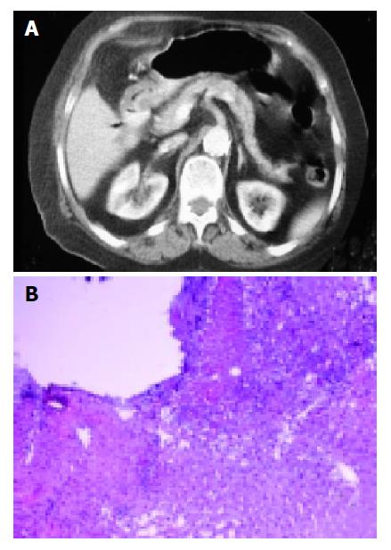
Figure 1 A: CT demonstrates fibrofatty in the liver next to liver; B: Endoscopic biopsy material shows deep ulceration of mucosa of duodenum and attaching liver tissue (hematoxylin-eosin ×200).
- Citation: Kircali B, Saricam T, Ozakyol A, Vardareli E. Endoscopic biopsy: Duodenal ulcer penetrating into liver. World J Gastroenterol 2005; 11(21): 3329-3329
- URL: https://www.wjgnet.com/1007-9327/full/v11/i21/3329.htm
- DOI: https://dx.doi.org/10.3748/wjg.v11.i21.3329


