©2005 Baishideng Publishing Group Inc.
World J Gastroenterol. Apr 21, 2005; 11(15): 2324-2329
Published online Apr 21, 2005. doi: 10.3748/wjg.v11.i15.2324
Published online Apr 21, 2005. doi: 10.3748/wjg.v11.i15.2324
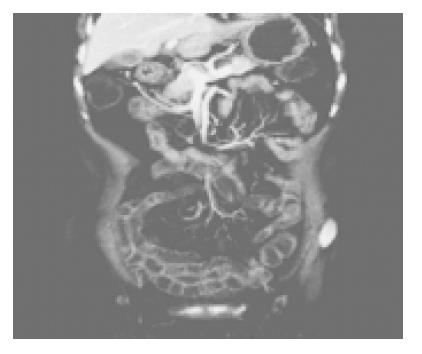
Figure 1 Coronal section shows adequate small bowel luminal distention, uniform wall enhancement and normal superior mesenteric vessels and their branching.
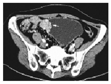
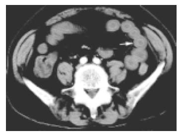
Figure 2 Axial CT-E image of lower abdomen shows an irregular mass at the obstruction point (arrow) which is supposed to be at the ileum.
Figure 3 Value of coronal images.
A: Cronal projection of the same patiet as fig 2 vividly displays the enhanced mass (arrow) at the dilated and elongated proximal jejunum; B: Coronal projection of a jejunal GIST clearly exhibits subserously located, markedly enhanced the mass (thin arrow) and its feeding artery and draining vein (thick arrow). C: Coronal projection of a MALT lymphoma shows aneurysmal dilatation of a long segment of thickened ileal loop that cannot be seperated from the bladder and multiple mesenteric nodes. D: Coronal projection of a patient with Crohn’s disease shows segmental ileal wall thickening, mucosal enhancement, prominent perienteric vasculacture and cutaneous orificium fistulae (arrow).
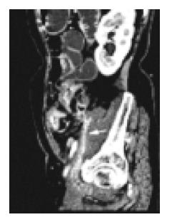
Figure 4 Sagittal projection of the same patient as figure 3D shows a markedly enhanced enterocutaneous fistula (arrow).
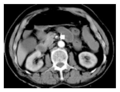
Figure 5 Axial section of an acute mensenteric ischemia caused by thrombosis shows an filling defect in SMA (arrow).
Figure 6 Lower section of the same patient as Figure 5 shows small bowel wall thickening with mural stratification.
- Citation: Zhang LH, Zhang SZ, Hu HJ, Gao M, Zhang M, Cao Q, Zhang QW. Multi-detector CT enterography with iso-osmotic mannitol as oral contrast for detecting small bowel disease. World J Gastroenterol 2005; 11(15): 2324-2329
- URL: https://www.wjgnet.com/1007-9327/full/v11/i15/2324.htm
- DOI: https://dx.doi.org/10.3748/wjg.v11.i15.2324


