©2005 Baishideng Publishing Group Inc.
World J Gastroenterol. Apr 21, 2005; 11(15): 2224-2229
Published online Apr 21, 2005. doi: 10.3748/wjg.v11.i15.2224
Published online Apr 21, 2005. doi: 10.3748/wjg.v11.i15.2224
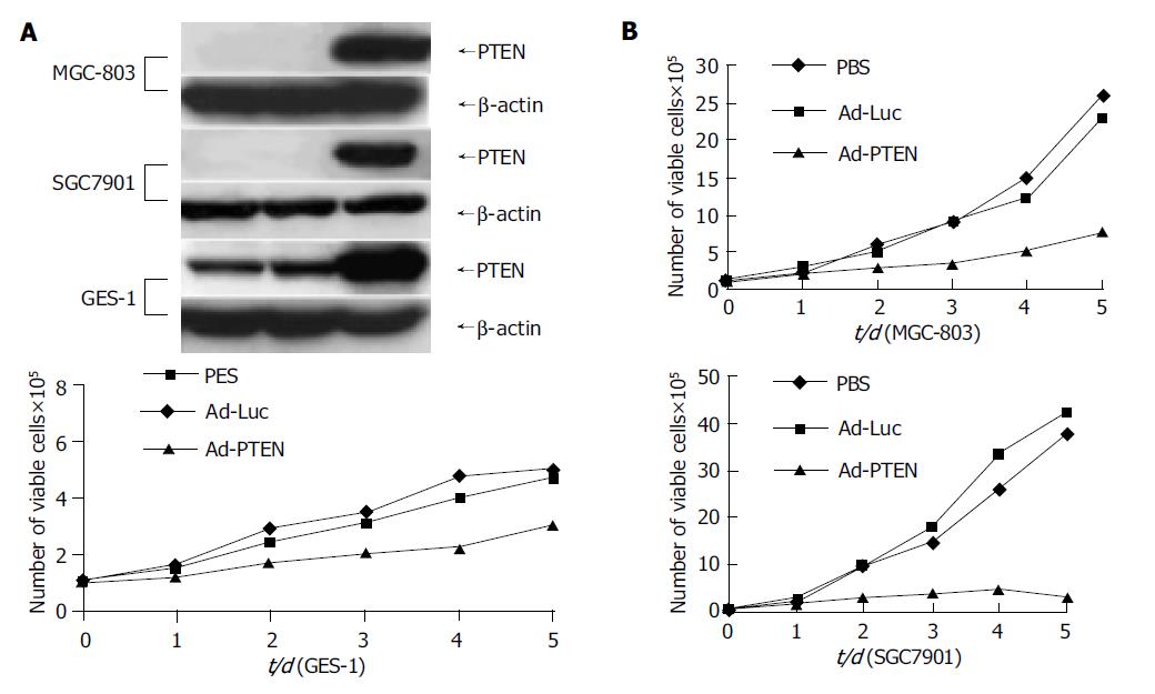
Figure 1 Inhibition of cell proliferation in gastric cancer cells due to overexpression of PTEN.
A: Cells analyzed for PTEN protein expression by Western blot analysis; B: Measurement of cell proliferation as determined by viability assay on d 1, 2, 3, 4, and 5.
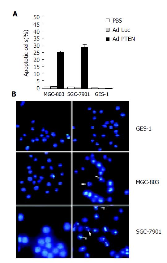
Figure 2 Induction of apoptosis due to overexpression of PTEN.
A: Tumor cells (MGC-803, SGC-7901) and normal cells (GES-1) treated with PBS, Ad-Luc, or Ad-PTEN and harvested at 72 h after treatment; B: Analysis of tumor cells (MGC-803, SGC-7901) and normal cells (GES-1) by Hoechst 33258 staining 72 h after treatment with Ad-Luc and Ad-PTEN. Arrows indicate apoptotic cells. Magnification ×40 for all cell lines.
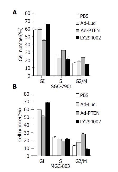
Figure 3 Induction of G2/M cell-cycle arrest due to overexpression of PTEN.
(A: SGC-7901 cells; B: MGC-803 cells.) Bars denote standard error (SE).
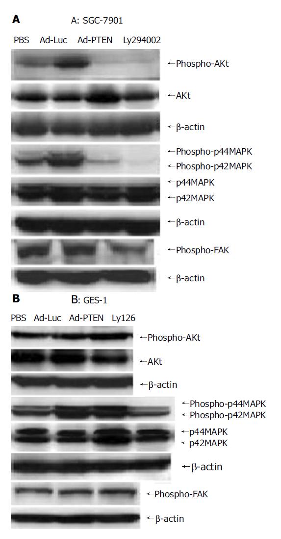
Figure 4 Signaling pathways regulated by PTEN overexpression.
A: Downregulation of phospho-Akt, phospho-FAK, and inhibition of phospho-p44/42MAPK in SGC-7901 cells after Ad-PTEN treatment compared to Ad-Luc- and PBS-treated cells; B: No change in the expression levels of phospho-Akt, phospho-FAK, and phospho-p44/42MAPK observed in GES-1 cells after Ad-PTEN treatment compared to Ad-Luc- and PBS-treated cells.
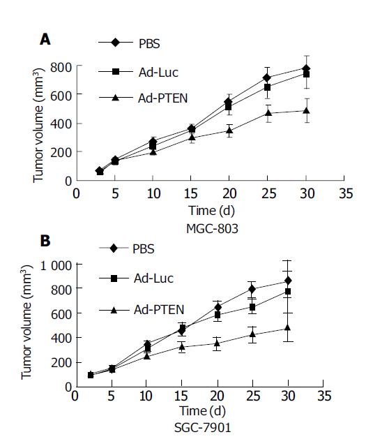
Figure 5 Therapeutic effect of Ad-PTEN on subcutaneous human gastric tumor xenografts.
Bars represent standard error (SE). A: MGC-803 cells; B: SGC-7901 cells.
- Citation: Hang Y, Zheng YC, Cao Y, Li QS, Sui YJ. Suppression of gastric cancer growth by adenovirus-mediated transfer of the PTEN gene. World J Gastroenterol 2005; 11(15): 2224-2229
- URL: https://www.wjgnet.com/1007-9327/full/v11/i15/2224.htm
- DOI: https://dx.doi.org/10.3748/wjg.v11.i15.2224


