Copyright
©2005 Baishideng Publishing Group Inc.
World J Gastroenterol. Apr 7, 2005; 11(13): 2013-2015
Published online Apr 7, 2005. doi: 10.3748/wjg.v11.i13.2013
Published online Apr 7, 2005. doi: 10.3748/wjg.v11.i13.2013
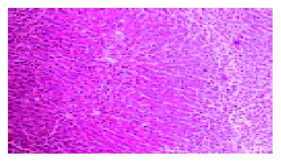
Figure 1 Histological feature of liver before biliary decompression (HE, ×70).
The structure of the interlobular biliary duct disappear, infiltrated with lymphocytes and neutrophil. Liver lobular is surrounded by dense fibrosis, with the presence of septa.
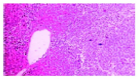
Figure 2 Histological feature of liver after biliary decompression of same patient (HE, ×70).
The structure of the interlobular biliary duct is still preserved. The infiltration with lymphocytes and neutrophil decreases.
- Citation: Chen ZB, Zheng SS, Hu GZ, Gao Y, Ding CY, Zhang Y, Zhao XH, Ni LM. Liver fibrosis caused by choledocholith to regress after biliary drainage. World J Gastroenterol 2005; 11(13): 2013-2015
- URL: https://www.wjgnet.com/1007-9327/full/v11/i13/2013.htm
- DOI: https://dx.doi.org/10.3748/wjg.v11.i13.2013


