Copyright
©2005 Baishideng Publishing Group Inc.
World J Gastroenterol. Mar 14, 2005; 11(10): 1439-1444
Published online Mar 14, 2005. doi: 10.3748/wjg.v11.i10.1439
Published online Mar 14, 2005. doi: 10.3748/wjg.v11.i10.1439
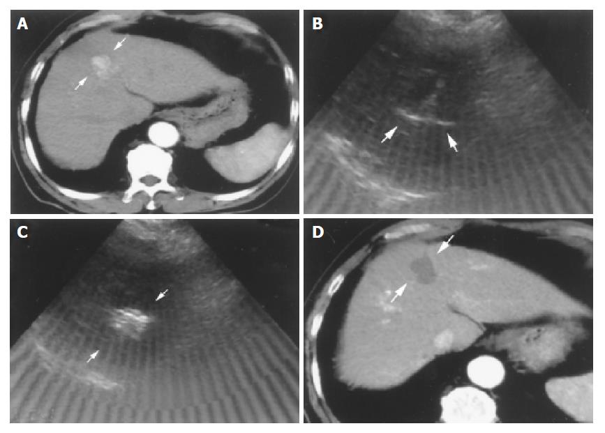
Figure 1 Successful RFA in an 82-year-old male patient with HCC.
A: Tri-phase CT scan showed an enhanced nodule in hepatic arterial phase about 2.8 cm in size before RFA (arrows); B: Radiofrequency ablation was performed in one session. The needle was introduced into the lower part of the tumor first with fully expanded tines under real-time ultrasound-guidance (arrows); C: Complete ablation after 2 ablative courses within 37 min was done. The tumor showed hyperechoic signals after RFA under ultrasound (arrows); D: Tri-phase CT scan showed complete necrosis of this tumor without enhancement in hepatic arterial phase one month after RFA (arrows).
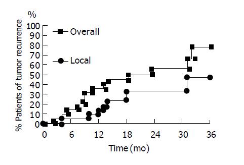
Figure 2 Accumulated tumor recurrence curves of 22 patients with small HCC after RFA.
Local tumor recurrence (triangle line) and overall tumor recurrence (including local tumor recurrence, new tumor formation and both) (dot line) are shown.
- Citation: Yu HC, Cheng JS, Lai KH, Lin CP, Lo GH, Lin CK, Hsu PI, Chan HH, Lo CC, Tsai WL, Chen WC. Factors for early tumor recurrence of single small hepatocellular carcinoma after percutaneous radiofrequency ablation therapy. World J Gastroenterol 2005; 11(10): 1439-1444
- URL: https://www.wjgnet.com/1007-9327/full/v11/i10/1439.htm
- DOI: https://dx.doi.org/10.3748/wjg.v11.i10.1439


