Copyright
©The Author(s) 2004.
World J Gastroenterol. Apr 1, 2004; 10(7): 977-984
Published online Apr 1, 2004. doi: 10.3748/wjg.v10.i7.977
Published online Apr 1, 2004. doi: 10.3748/wjg.v10.i7.977
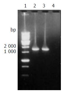
Figure 1 The target fragment of ureB gene amplified from H pylori strain Y06 DNA.
Lane 1, 1 kb DNA marker (BBST); lanes 2 and 3, the target amplification fragment of ureB gene from genomic DNA of H pylori strain Y06; lane 4: blank control.
Figure 2 Homology comparison of the cloned and reported H pylori ureB nucleotide sequences.
(1)-(3): the reported sequences from GenBank (No. NC000915, strain 26695; No. M60398, X57132; No. AB032429); (4): the sequencing result of H pylori strain Y06 ureB gene. Underlined areas indicate the positions of primer sequences.
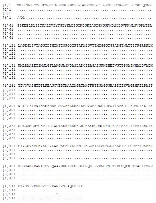
Figure 3 Homology comparison of the putative amino acid sequences of the cloned and reported H pylori ureB gene.
[1]-[3]: the reported sequences from GenBank (No. NC000915, strain 26695; No. M60398, X57132; No. AB032429); [4]: the putative amino acid sequence of the cloned H pylori strain Y06 ureB gene.
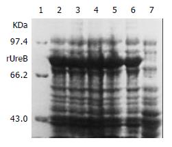
Figure 4 Expression of rUreB induced by IPTG at different concentrations.
Lane 1, molecular mass marker; lanes 2-4, IPTG at 1.0, 0.5, and 0.1 mmol/L, respectively; lanes 5 and 6, bacterial precipitate and supernatant with IPTG at 1 mmol/L , respectively; lane 7, control.
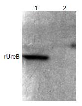
Figure 5 Western blot result of commercial rabbit antibodies against the whole cell of H pylori and rUreB.
Lane 1, rUreB; lane 2, blank.
- Citation: Mao YF, Yan J. Construction of prokaryotic expression system of ureB gene from a clinical Helicobacter pylori strain and identification of the recombinant protein immunity. World J Gastroenterol 2004; 10(7): 977-984
- URL: https://www.wjgnet.com/1007-9327/full/v10/i7/977.htm
- DOI: https://dx.doi.org/10.3748/wjg.v10.i7.977


