Copyright
©The Author(s) 2004.
World J Gastroenterol. Apr 1, 2004; 10(7): 954-958
Published online Apr 1, 2004. doi: 10.3748/wjg.v10.i7.954
Published online Apr 1, 2004. doi: 10.3748/wjg.v10.i7.954
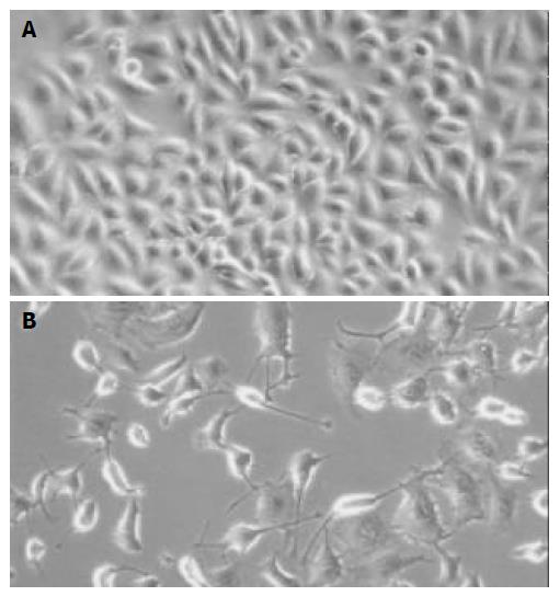
Figure 1 Detection of cell morphology by phase-contrast microscopy ( × 200).
A: SMMC-7721 cells, B: SMMC-7721 cells treated with 10 mmol/L of HMBA for 72 h.
Figure 2 Detection of apoptotic morphology in SMMC-7721 cells treated with 10 mmol/L of HMBA for 72 h ( × 400).
SMMC-7721 cells displayed fragmented nuclei and apoptotic bodies (B, C) after treatment with 10 mmol/L of HMBA for 72 h, while the untreated cells did not show these apoptotic characteristics (A).
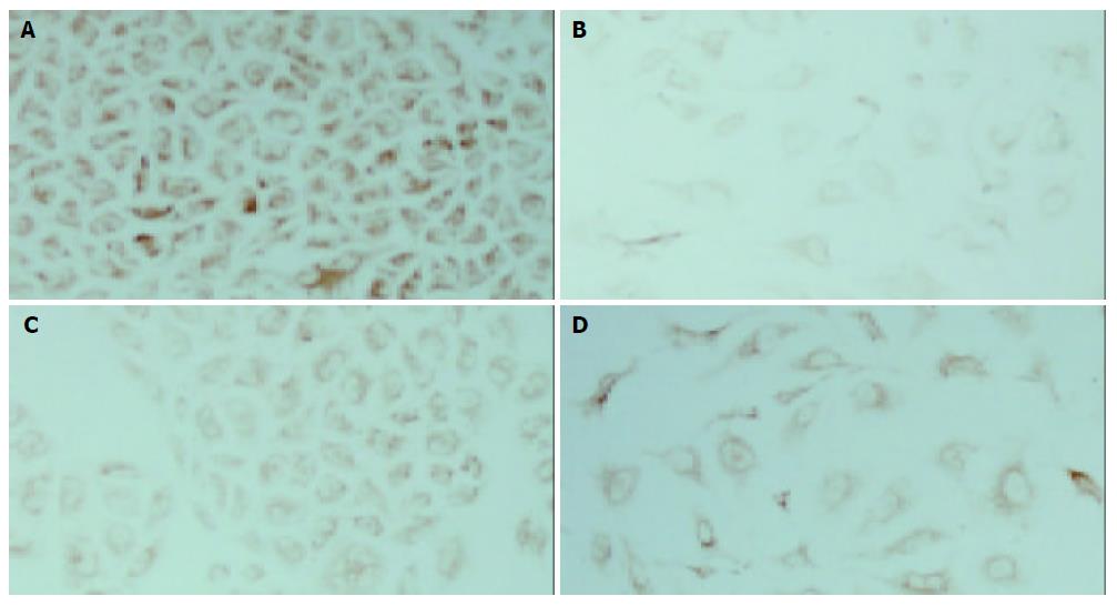
Figure 3 Immunocytochemical analysis of Bcl-2 and Bax expression in SMMC-7721 cells treated with 10 mmol/L of HMBA for 72 h (×200).
A: The high level of Bcl-2 protein in SMMC-7721 cells, B: The decreased level of Bcl-2 protein in the cells treated with 10 mmol/L of HMBA, C: The low level of Bax protein in SMMC-7721 cells, D: The slightly increased level of Bax protein in the cells treated with 10 mmol/L of HMBA.
- Citation: Ouyang GL, Cai QF, Liu M, Chen RC, Huang Z, Jiang RS, Chen F, Hong SG, Bao SD. Growth arrest and apoptosis of human hepatocellular carcinoma cells induced by hexamethylene bisacetamide. World J Gastroenterol 2004; 10(7): 954-958
- URL: https://www.wjgnet.com/1007-9327/full/v10/i7/954.htm
- DOI: https://dx.doi.org/10.3748/wjg.v10.i7.954


