Copyright
©The Author(s) 2004.
World J Gastroenterol. Apr 1, 2004; 10(7): 1065-1068
Published online Apr 1, 2004. doi: 10.3748/wjg.v10.i7.1065
Published online Apr 1, 2004. doi: 10.3748/wjg.v10.i7.1065
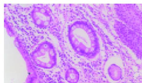
Figure 1 Goblet cells with HE stain (10 × 10).
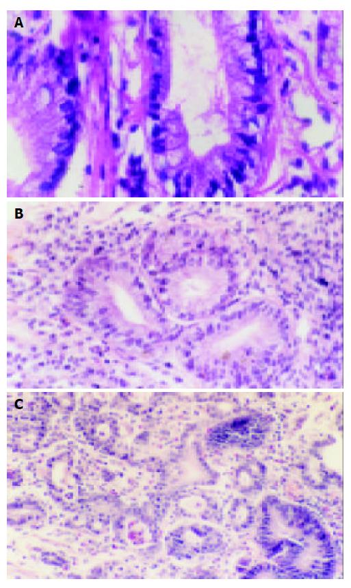
Figure 2 A: Barrett’s esophagus with LGD (10 × 40), B: Barrett’s esophagus with LGD (10 × 10), C: Barrett’s esophagus with HGD and carcinogenesis (top left corner) (10 × 10).
- Citation: Zhang J, Chen XL, Wang KM, Guo XD, Zuo AL, Gong J. Barrett’s esophagus and its correlation with gastroesophageal reflux in Chinese. World J Gastroenterol 2004; 10(7): 1065-1068
- URL: https://www.wjgnet.com/1007-9327/full/v10/i7/1065.htm
- DOI: https://dx.doi.org/10.3748/wjg.v10.i7.1065


