Copyright
©The Author(s) 2004.
World J Gastroenterol. Apr 1, 2004; 10(7): 1047-1051
Published online Apr 1, 2004. doi: 10.3748/wjg.v10.i7.1047
Published online Apr 1, 2004. doi: 10.3748/wjg.v10.i7.1047
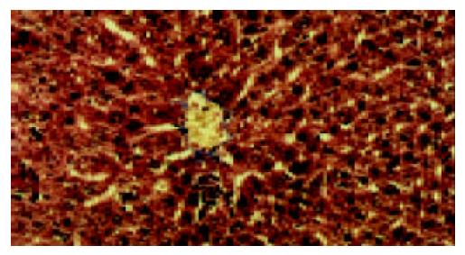
Figure 1 Liver tissue from control group showed normal lobular architecture and a normal distribution of collagen with a thin rim around central veins.
Masson × 200.
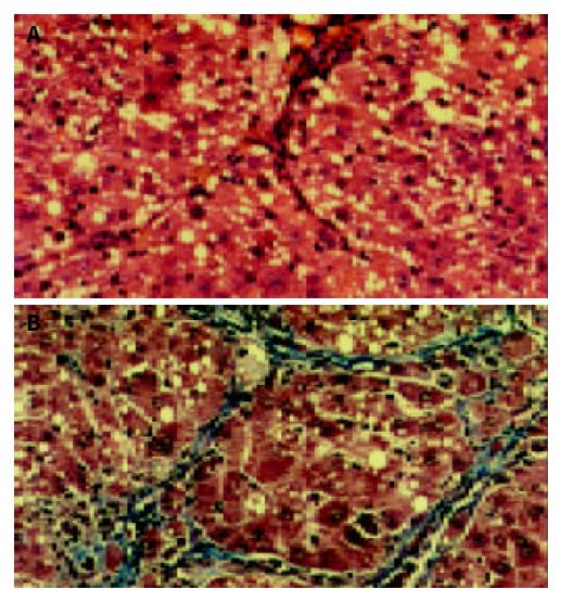
Figure 2 A: Liver tissue from model group showed disorderly hepatocyte cords, severe fatty degeneration, spotty or focal necrosis and infiltration of inflammatory cells.
HE × 200. B: Liver tissue from model group showed collagen deposition extending from central veins or portal tracts, with thick or thin fibrotic septa and pseudolobuli formation. Masson × 200.
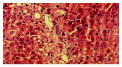
Figure 3 Liver tissue from PII group showed apparent amelio-ration of hepatocyte degeneration, necrosis and infiltration of inflammatory cells.
HE × 200.
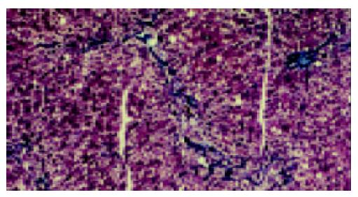
Figure 4 Liver tissue from PII group showed marked reduction in collagen deposition with no obvious pseudolobuli formation.
Masson × 100.
- Citation: Yuan GJ, Zhang ML, Gong ZJ. Effects of PPARg agonist pioglitazone on rat hepatic fibrosis. World J Gastroenterol 2004; 10(7): 1047-1051
- URL: https://www.wjgnet.com/1007-9327/full/v10/i7/1047.htm
- DOI: https://dx.doi.org/10.3748/wjg.v10.i7.1047


