Copyright
©The Author(s) 2004.
World J Gastroenterol. Feb 15, 2004; 10(4): 545-549
Published online Feb 15, 2004. doi: 10.3748/wjg.v10.i4.545
Published online Feb 15, 2004. doi: 10.3748/wjg.v10.i4.545
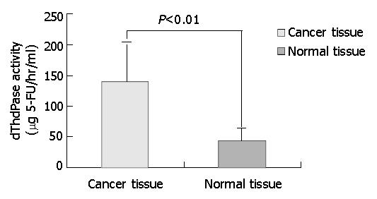
Figure 1 dThdPase activity analysis in 27 specimens of colorectal carcinoma.
The dThdPase Activity was 139.7 ± 42.2 (μg 5-FU/hr/ml) in cancer tissues, significantly higher than 61.5 ± 21.4 (μg 5-FU/hr/ml) in adjacent normal tissue (P < 0.01).
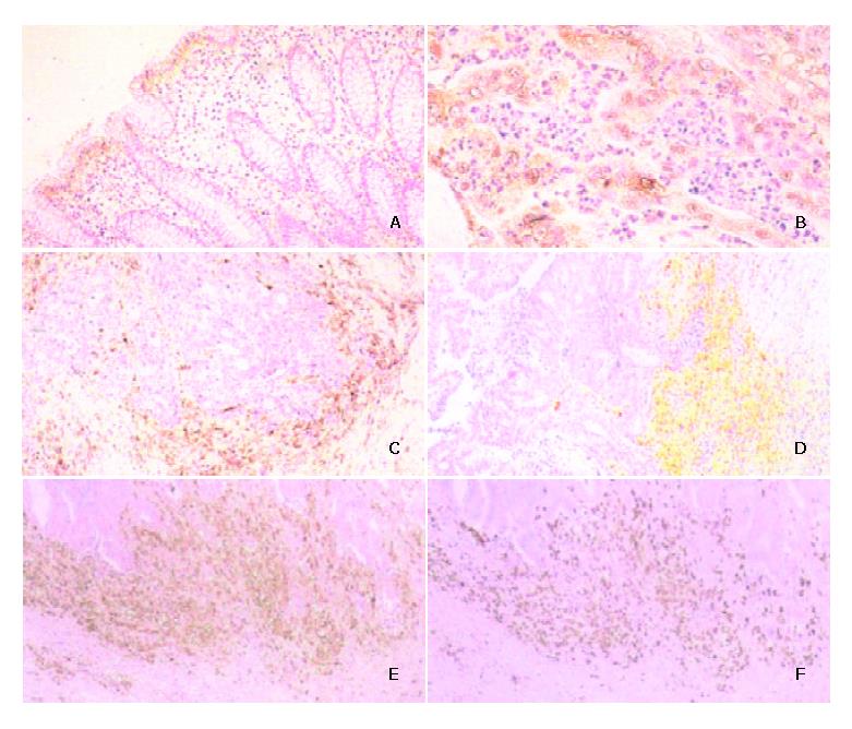
Figure 2 Immunohistochemical staining in colorectal cancer tissues.
A: In normal colon mucosa, epithelial cells at the surface are weakly positive for dThdPase and few stromal cells are stained. B: In 3 of the 39 cases examined, cancer cells are positive for dThdPase. C: In most cases of colorectal cancers examined, stromal cells around the cancer nests are positive for dThdPase, while cancer cells are negative. D: In some cases, dThdPase-positive stromal cells are along the invasive margin of cancer, cancer cells are negative for dThdPase. E: Immunohistochemical staining for dThdPase and F: for CD68 in the corresponding area of colorectal cancer tissue. dThdPase-positive stromal cells are observed densely along the invasive margin of cancer. The distribution pattern of dThdPase-positive cells (E) is similar to that of CD68-positive cells (F).
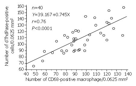
Figure 3 Correlation between number of CD68-positive mac-rophages and dThdPase-positive stromal cells in 40 cases of colorectal cancer.
The significant correlation is judged by Spearman’s coeffecient rank test (r = 0.76, P < 0.0001).
- Citation: Zhang JM, Mizoi T, Shiiba KI, Sasaki I, Matsuno S. Expression of thymidine phosphorylase by macrophages in colorectal cancer tissues. World J Gastroenterol 2004; 10(4): 545-549
- URL: https://www.wjgnet.com/1007-9327/full/v10/i4/545.htm
- DOI: https://dx.doi.org/10.3748/wjg.v10.i4.545


