Copyright
©The Author(s) 2004.
World J Gastroenterol. Feb 1, 2004; 10(3): 342-347
Published online Feb 1, 2004. doi: 10.3748/wjg.v10.i3.342
Published online Feb 1, 2004. doi: 10.3748/wjg.v10.i3.342
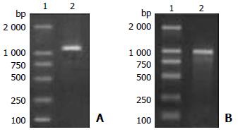
Figure 1 Agarose gel electrophoresis of PCR product.
A: hMC-CP cDNA (lane 1: DNA molecular marker; lane 2: hMC-CP cDNA). B: coding region of mature human colon MC-CP cDNA (lane 1: DNA molecular marker; lane 2: PCR product of colon MC-CP cDNA).
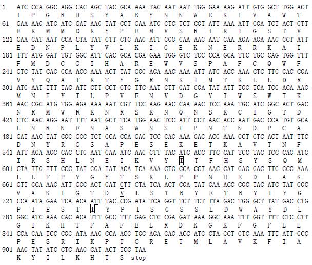
Figure 2 Nucleotide sequence and deduced amino acid sequences of mature human colon MC-CP.
The nucleotide variations and amino acid substitutions different from the skin MC-CP are underlined and boxed, respectly.
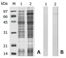
Figure 3 Analysis of recombinant proteins expressed in E.
coli. A: SDS-PAGE. B: Western blots. M: molecular mass markers; lane 1: without IPTG induction; lane 2: with IPTG induction.
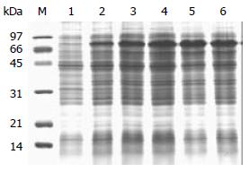
Figure 4 SDS-PAGE analysis of time course of recombinant proteins expressed in E.
coli. lane 1: before induction; lane 2: 8 h after induction; lane 3: 16 h after induction; lane 4: 24 h after induction; lane 5: 32 h after induction.
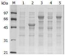
Figure 5 SDS-PAGE analysis of rhMC-CP expressed in E.
coli TB1 cells. M: molecular weight markers; lane 1: total cellular protein of E.coli TB1 cells without IPTG induction; lane 2: total cellular protein of E.coli TB1 cells with IPTG induction (control vector); lane 3: total cellular protein of E.coli TB1 cells with IPTG induction; lane 4: soluble fraction of cell lysate from E.coli TB1 with IPTG induction; lane 5: precipitated fraction of cell lysate from E.coli TB1 with IPTG induction.
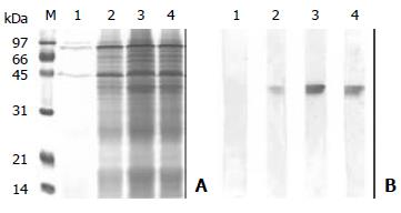
Figure 6 Induction of recombinant HMC-CP expressed in P.
pastoris. A: secreted proteins analyzed by SDS-PAGE. B: Western blot analysis of secreted proteins with HMC-specific monoclonal antibody, clone CA5. M: molecular weight markers; lane 1: 0 h; lane 2: 24 h; lane 3: 48 h; lane 4: 72 h.
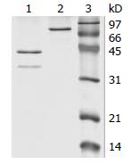
Figure 7 SDS-PAGE analysis of fusion protein cleavage (lane 1: cleaved by factor Xa; lane 2: uncleaved by factor Xa; lane 3: molecular weight marker).
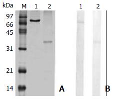
Figure 8 Analysis of purified recombinant protein.
A: SDS-PAGE analysis of purified recombinant protein. B: Western blot analysis of purified fusion protein with CA5. Lane 1: purified fusion protein after MBP affinity chromatography; lane 2: pu-rified recombinant hMC-CP after heparin agarose affinity chromatography.
- Citation: Chen ZQ, He SH. Cloning and expression of human colon mast cell carboxypeptidase. World J Gastroenterol 2004; 10(3): 342-347
- URL: https://www.wjgnet.com/1007-9327/full/v10/i3/342.htm
- DOI: https://dx.doi.org/10.3748/wjg.v10.i3.342


