©The Author(s) 2004.
World J Gastroenterol. Dec 1, 2004; 10(23): 3534-3536
Published online Dec 1, 2004. doi: 10.3748/wjg.v10.i23.3534
Published online Dec 1, 2004. doi: 10.3748/wjg.v10.i23.3534
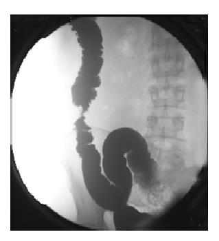
Figure 1 Stenosis at the segment of descending colon, less than 5 mm in diameter.
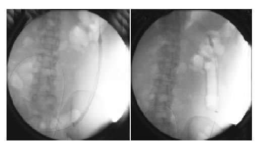
Figure 2 The first stent was appropriately positioned and deployed by withdrawing the enveloping membrane under fluoroscopic control to ensure that the lesion was adequately covered, thus relieving the obstruction.
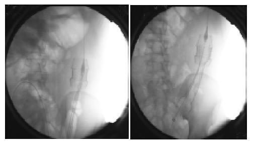
Figure 3 Clinical symptoms of obstruction recurred the next day.
Anteroposterior radiographs showed the stent migrated above the lesion and a second stent was needed. Second stent was deployed with the same stent placement techniques through the lumen of the first stent.
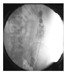
Figure 4 Radiograph obtained after a water-soluble enema on the day after the second stent was deployed shows that the stents expanded to provide an adequate lumen.
- Citation: Guan YS, Sun L, Li X, Zheng XH. Successful management of a benign anastomotic colonic stricture with self-expanding metallic stents: A case report. World J Gastroenterol 2004; 10(23): 3534-3536
- URL: https://www.wjgnet.com/1007-9327/full/v10/i23/3534.htm
- DOI: https://dx.doi.org/10.3748/wjg.v10.i23.3534


