Copyright
©The Author(s) 2004.
World J Gastroenterol. Nov 1, 2004; 10(21): 3171-3174
Published online Nov 1, 2004. doi: 10.3748/wjg.v10.i21.3171
Published online Nov 1, 2004. doi: 10.3748/wjg.v10.i21.3171
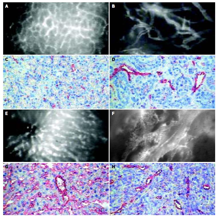
Figure 1 Microangioarchtecture investigated by intravital microscopy and immunohistochemical staining of endothelium of normal pancreas (A,B), liver (E,F), pancreatic (C,D) and hepatocellular (G,H) carcinoma: The microvascular system in healthy pancreas showed a dense network mainly consisting of capillaries in normal pancreas (A) and sinusoids in the liver (E).
Both pancreatic (B) and hepatocellular (F) carcinomas showed a chaotic angioarchitecture with irregular diameter of blood vessels. Either intravital microscopy or immunohistochemical staining of endothelium turn up lower density and higher diameters of microvessels in both tumor types than in corresponding normal tissue (bar 50 µm).
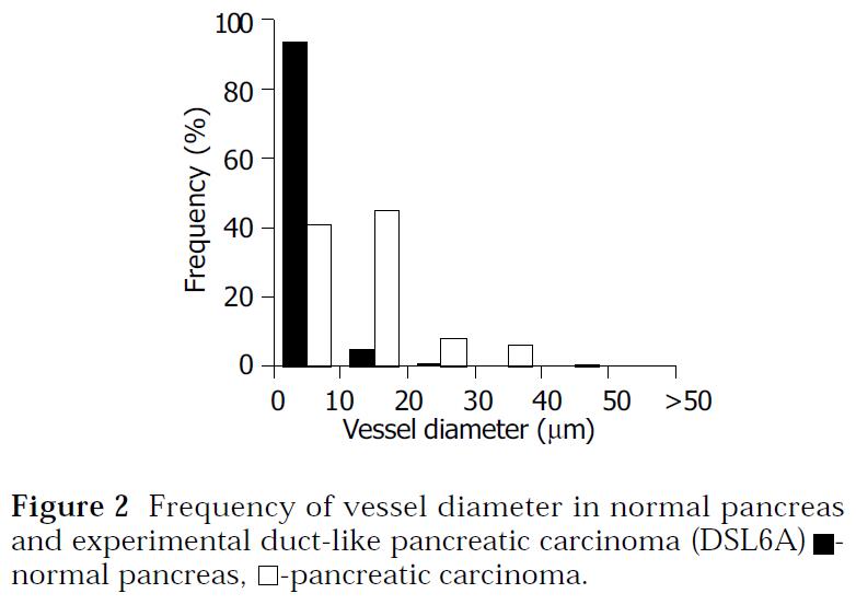
Figure 2 Frequency of vessel diameter in normal pancreas and experimental duct-like pancreatic carcinoma (DSL6A) ■- normal pancreas, □-pancreatic carcinoma.
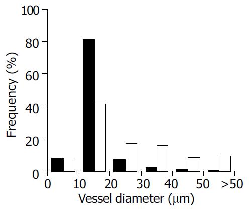
Figure 3 Frequency of vessel diameter in normal liver and experimental hepatocellular carcinoma (Morris-hepatoma) ■- normal liver, □-hepatocellular carcinoma.
- Citation: Ryschich E, Schmidt E, Maksan SM, Klar E, Schmidt J. Expansion of endothelial surface by an increase of vessel diameter during tumor angiogenesis in experimental hepatocellular and pancreatic cancer. World J Gastroenterol 2004; 10(21): 3171-3174
- URL: https://www.wjgnet.com/1007-9327/full/v10/i21/3171.htm
- DOI: https://dx.doi.org/10.3748/wjg.v10.i21.3171


