©The Author(s) 2004.
World J Gastroenterol. Nov 1, 2004; 10(21): 3118-3121
Published online Nov 1, 2004. doi: 10.3748/wjg.v10.i21.3118
Published online Nov 1, 2004. doi: 10.3748/wjg.v10.i21.3118
Figure 1 Images of multi-phasic CTAP and CTHA.
A: A nodular perfusion defect of HCC on CTAP image. B: A small round enhancement nodule of HCC on early-phasic CTHA image. C: Rim enhancement of HCC on delayed-phasic CTHA image obtained 2 min later.
Figure 2 HCC on early-phasic CTAP and late-phasic CTHA.
A: HCC shown as a well-fined hypodense nodule on early-phasic CTAP image. B: HCC demonstrated as rim enhancement on late-phasic CTHA image. C: HCC demonstrated as irregular enhance-ment on CTHA image.
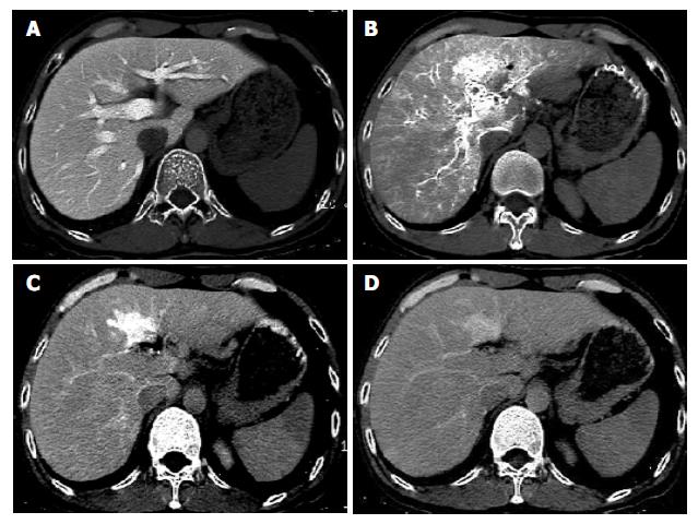
Figure 3 Pseudolesions on CTAP and CTHA.
A: No lesion demonstrated on CTAP. B: Pseudolesion shown as an irregular enhancement focus on early-phasic CTHA. C: Change in shape and size of pseudolesion on late-phasic CTAP. D: Pseudolesion demonstrated as isodensity to liver tissue on delayed-CTHA.
Figure 4 Hemangiomas on CTAP and CTHA.
A: A small hemangioma in segment II of left lobe as a perfusion defect nodule on CTAP. B: Lesion demonstrated as a punctulate, peripheral enhancement focus on early-phasic CTHA. C: Hemangioma demon-strated as hyperdensity to liver tissue on delayed-CTHA.
- Citation: Li L, Liu LZ, Xie ZM, Mo YX, Zheng L, Ruan CM, Chen L, Wu PH. Multi-phasic CT arterial portography and CT hepatic arteriography improving the accuracy of liver cancer detection. World J Gastroenterol 2004; 10(21): 3118-3121
- URL: https://www.wjgnet.com/1007-9327/full/v10/i21/3118.htm
- DOI: https://dx.doi.org/10.3748/wjg.v10.i21.3118


