Copyright
©The Author(s) 2004.
World J Gastroenterol. Sep 1, 2004; 10(17): 2613-2615
Published online Sep 1, 2004. doi: 10.3748/wjg.v10.i17.2613
Published online Sep 1, 2004. doi: 10.3748/wjg.v10.i17.2613
Figure 1 CT scans of splenic hamartoma.
A: Homogeneous mild hypodensity lesion within the spleen found by unenhanced CT scan, B: Mild-homogeneous enhancement of the mass found by enhanced CT scan on hepatic artery phase, C: Isodense tumor with normal spleen on delayed enhanced CT images.
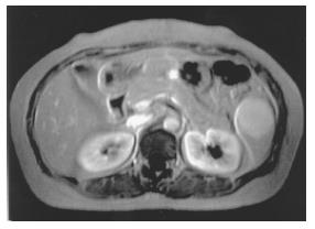
Figure 2 MR images of splenic hamartoma.
A: Round-like mass with isointensity relative to the spleen on MR T1-weighted images, B: Mild-hypointense mass on MR T2-weighted images, C: Diffuse heterogeneous enhancement of mass by enhanced MR scan on early portal venous phase.
Figure 3 Obviously hyperintense tumor with normal spleen on delayed enhanced MR images.
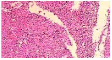
Figure 4 Red marrow tissue and some blood sinusoid structures with lymphocytes and macrophages in hamartoma.
Fibrosis was remarkable. (HE, original magnification × 100).
- Citation: Yu RS, Zhang SZ, Hua JM. Imaging findings of splenic hamartoma. World J Gastroenterol 2004; 10(17): 2613-2615
- URL: https://www.wjgnet.com/1007-9327/full/v10/i17/2613.htm
- DOI: https://dx.doi.org/10.3748/wjg.v10.i17.2613


