Copyright
©The Author(s) 2004.
World J Gastroenterol. Aug 15, 2004; 10(16): 2455-2456
Published online Aug 15, 2004. doi: 10.3748/wjg.v10.i16.2455
Published online Aug 15, 2004. doi: 10.3748/wjg.v10.i16.2455
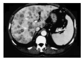
Figure 1 Contrast-enhanced abdominal CT scan (portal phase): multiple lesions appearing hypodense compared with normal liver parenchyma and showing mild ring enhancement.
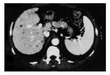
Figure 2 Contrast-enhanced abdominal CT scan (portal phase): after 46 d both number and volume of hepatic lesions were considerably decreased.
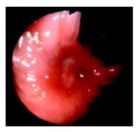
Figure 3 Endoscopic view of right colon showing an ulcer-ated lesion without signs of bleeding.
A series of biopsies of the ulcer revealed only necro-inflammatory cells.
- Citation: Granito A, Ballardini G, Fusconi M, Volta U, Muratori P, Sambri V, Battista G, Bianchi FB. A case of leptospirosis simulating colon cancer with liver metastases. World J Gastroenterol 2004; 10(16): 2455-2456
- URL: https://www.wjgnet.com/1007-9327/full/v10/i16/2455.htm
- DOI: https://dx.doi.org/10.3748/wjg.v10.i16.2455


