Copyright
©The Author(s) 2004.
World J Gastroenterol. Aug 1, 2004; 10(15): 2245-2249
Published online Aug 1, 2004. doi: 10.3748/wjg.v10.i15.2245
Published online Aug 1, 2004. doi: 10.3748/wjg.v10.i15.2245
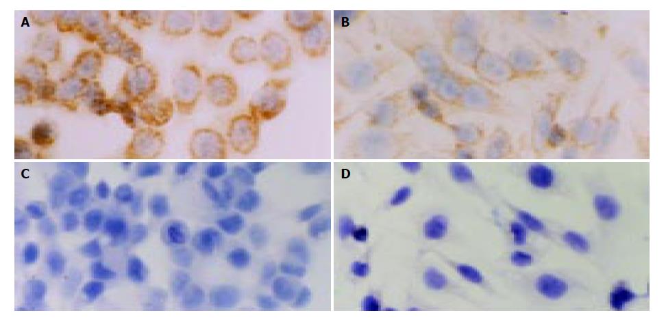
Figure 1 In situ hybridization detection of expression of KAI1 mRNA in colorectal carcinoma cell lines (Original magnification: × 400).
A: In HT29 cells, the positive expression (brown granule) was located in cytoplasm; B: In SW480 cells, the positive expression was located in cytoplasm; C: In SW620 cells, the expression of KAI1 mRNA was negative; D: In LoVo cells, the expression of KAI1 mRNA was negative.
Figure 2 Expression of KAI1 protein detected by immunohistochemistry in colorectal carcinoma cell lines (Original magnification: × 400).
A: In HT29 cells, the positive expression located in cytoplasm and membrane; B: In SW480 cells, the positive expression located in cytoplasm and membrane.
Figure 3 In situ hybridization and immunohistochemical detection of KAI1 mRNA and protein in colonic carcinoma (Original magnification: × 400).
A: In situ hybridization detection found that strong KAI1 mRNA staining was located in cytoplasm (brown granule); B: Detection by immunohistochemistry showed strong KAI1 protein staining was in cytoplasm and membrane.
- Citation: Wu DH, Liu L, Chen LH, Ding YQ. KAI1 gene expression in colonic carcinoma and its clinical significances. World J Gastroenterol 2004; 10(15): 2245-2249
- URL: https://www.wjgnet.com/1007-9327/full/v10/i15/2245.htm
- DOI: https://dx.doi.org/10.3748/wjg.v10.i15.2245


