Copyright
©The Author(s) 2004.
World J Gastroenterol. Jul 15, 2004; 10(14): 2091-2094
Published online Jul 15, 2004. doi: 10.3748/wjg.v10.i14.2091
Published online Jul 15, 2004. doi: 10.3748/wjg.v10.i14.2091
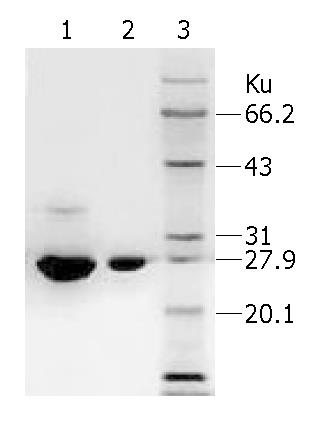
Figure 1 Analysis of EOT and TCS by SDS-PAGE.
Lane 1: EOT, Lane 2: TCS, Lane 3: Protein Marker.
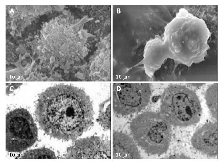
Figure 2 HeLa cells under SEM and TEM.
A: HeLa cells under SEM in control group. B: HeLa cells under SEM treated with EOT (100 mg/L) for 24 h. C: HeLa cells under TEM in control group. D: HeLa cells under TEM treated with EOT (100 mg/L) for 24 h.
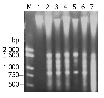
Figure 3 Analysis of DNA fragments of HeLa cells treated with EOT.
M: DNA marker, lane 1: normal control, lane 2: 500 mg/L for 24 h, lane 3: 100 mg/L for 24 h, lane 4: 50 mg/L for 48 h, lane 5: 50 mg/L for 24 h, lane 6: 100 mg/L for 48 h, lane 7: 500 mg/L for 48 h.
- Citation: Dou CM, Li JC. Effect of extracts of trichosanthes root tubers on HepA-H cells and HeLa cells. World J Gastroenterol 2004; 10(14): 2091-2094
- URL: https://www.wjgnet.com/1007-9327/full/v10/i14/2091.htm
- DOI: https://dx.doi.org/10.3748/wjg.v10.i14.2091


