Copyright
©The Author(s) 2004.
World J Gastroenterol. Jun 1, 2004; 10(11): 1574-1577
Published online Jun 1, 2004. doi: 10.3748/wjg.v10.i11.1574
Published online Jun 1, 2004. doi: 10.3748/wjg.v10.i11.1574
Figure 1 Massive rectal carcinoma in a 74 years old man.
VC, SSD displayed an irregular mass, but unable to determine circumfer-ential extent (A, B). MPR showed the mass with 1/4 circumference around rectal wall (C).
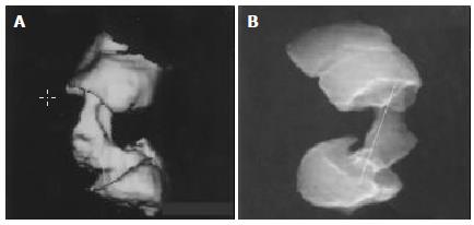
Figure 2 Infiltrative rectal carcinoma in a 46 years old man.
SSD disclosed the two ends of carcinoma and measured its length (A). Raysum manifested the two ends of carcinoma more clearly and measured its length more accurately than SSD (B).
Figure 3 Infiltrative rectal carcinoma in a 48 years old man.
VC demonstrated the carcinoma around rectal wall (A). Combination of coronal and sagittal images of MPR revealed an infiltrative carcinoma, but was not so obvious as VC (B, C).
- Citation: Luo MY, Shan H, Yao LQ, Zhou KR, Liang WW. Postprocessing techniques of CT colonography in detection of colorectal carcinoma. World J Gastroenterol 2004; 10(11): 1574-1577
- URL: https://www.wjgnet.com/1007-9327/full/v10/i11/1574.htm
- DOI: https://dx.doi.org/10.3748/wjg.v10.i11.1574


