Copyright
©The Author(s) 2020.
Artif Intell Med Imaging. Jun 28, 2020; 1(1): 40-49
Published online Jun 28, 2020. doi: 10.35711/aimi.v1.i1.40
Published online Jun 28, 2020. doi: 10.35711/aimi.v1.i1.40
Figure 1 Schematic diagram of acute peripancreatic fluid collections and magnetic resonance imaging of a patient.
A: Schematic diagram of acute peripancreatic fluid collections within 4 wk of onset of interstitial oedematous pancreatitis. The term “acute peripancreatic fluid collections” applies only to interstitial oedematous pancreatitis patients; B: A 66-year-old woman with interstitial oedematous pancreatitis. Magnetic resonance imaging axial T2WI image shows a homogeneous fluid finding (arrows) around the pancreas. APFCs: Acute peripancreatic fluid collections; P: Pancreas.
Figure 2 Schematic diagram of acute necrotic collections and computed tomography of a patient.
A: Schematic diagram of acute necrotic collections within 4 wk of onset of necrotizing pancreatitis. The term “acute necrotic collections” is diagnosed only in necrotizing pancreatitis patients; B: A 56-year-old man with necrotizing pancreatitis. Axial contrast-enhanced computed tomography image in the venous phase shows a large area of necrosis (asterisks) in the pancreatic body and tail; therefore, a lesser omental sac collection (arrows) should be diagnosed as acute necrotic collections. ANCs: Acute necrotic collections; P: Pancreas; N: Necrosis.
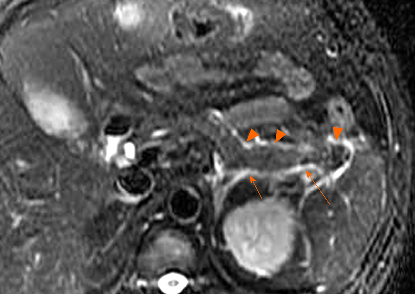
Figure 3 A 40-year-old woman with interstitial oedematous pancreatitis.
Magnetic resonance imaging axial T2WI image shows uniform linear liquid hyperintense (acute peripancreatic fluid collections) in the left pararenal anterior spaces (arrowheads) and retromesenteric plane (arrows).
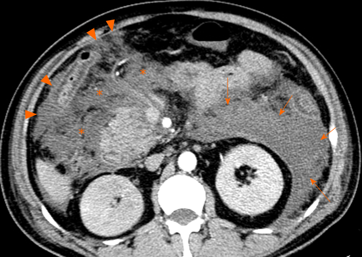
Figure 4 A 53-year-old man with necrotizing pancreatitis.
Axial contrast-enhanced computed tomography image in the venous phase shows extensive heterogeneous collections (acute necrotic collections) in the left pararenal anterior spaces (arrows) and the subperitoneal spaces/transverse mesentery areas (asterisks) as well as greater omentum zones (arrowheads).
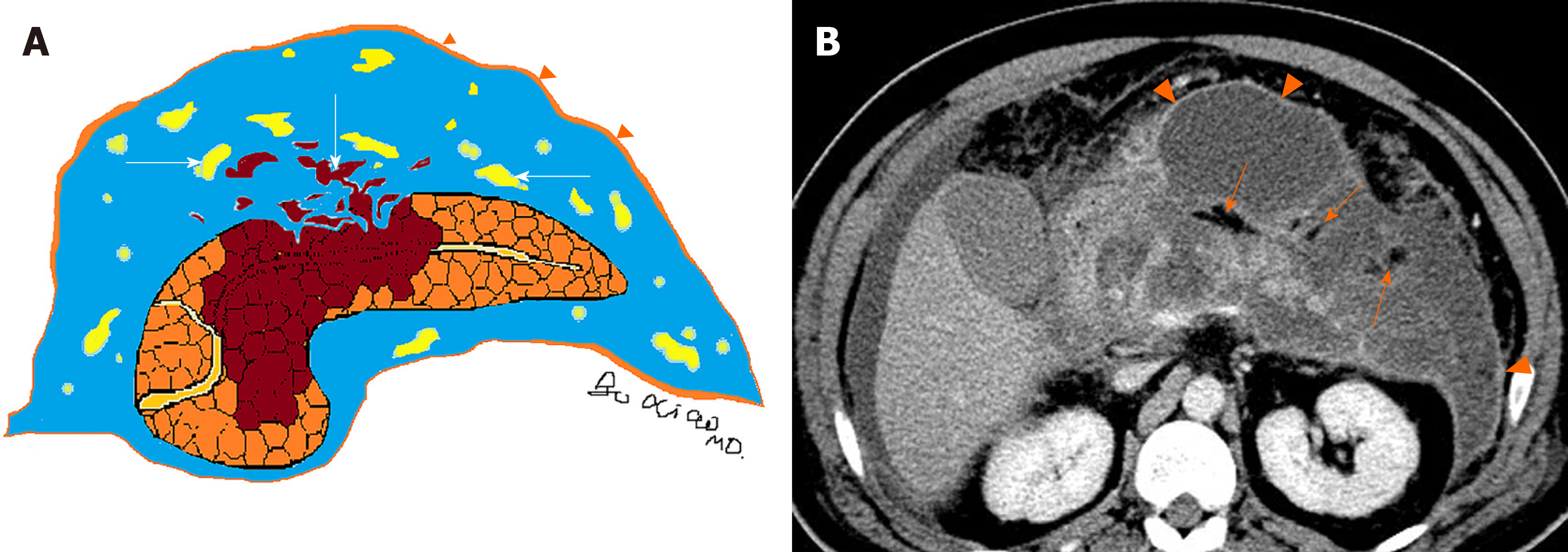
Figure 5 Schematic diagram of heterogeneous collections and computed tomography image of a patient.
A: Schematic diagram of heterogeneous collections (acute necrotic collections) secondary to necrotizing pancreatitis. With the prolongation of the disease course, immature poorly organized walls (arrowheads) can gradually form. There are two components within necrotic collections: fat fragments and necrotic parenchymal fragments (arrows); B: A 60-year-old man with necrotizing pancreatitis. Axial contrast-enhanced computed tomography image in the venous phase shows markedly hypodense fat globules (arrows) within acute necrotic collections as well as slight enhancement of capsule (arrowheads) of collections. P: Pancreas; N: Necrosis.
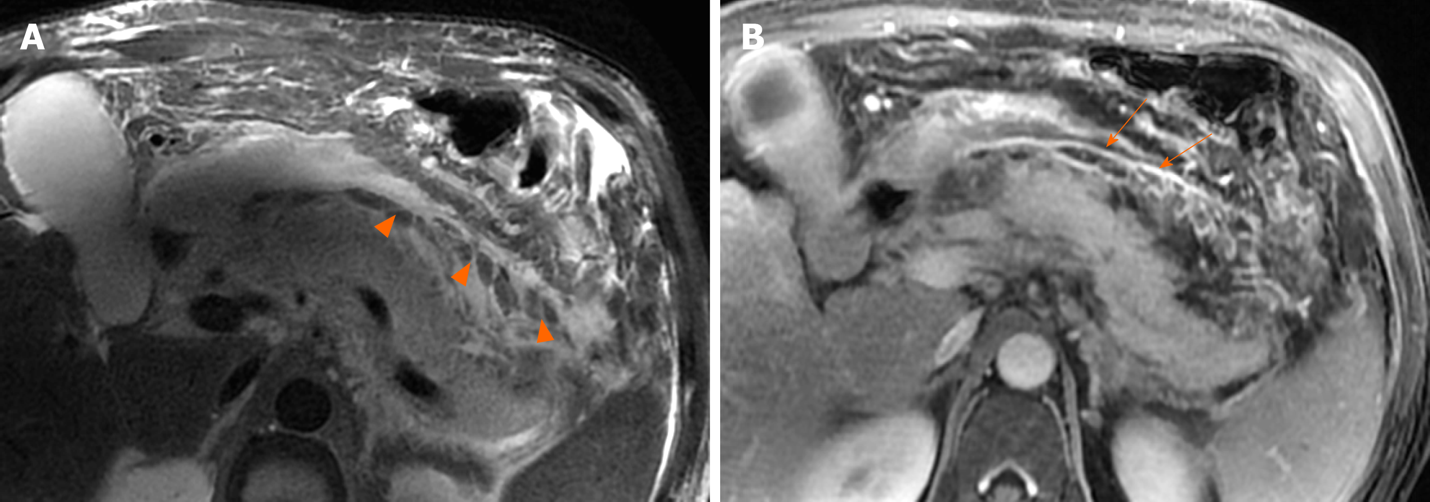
Figure 6 A 46-year-old man with necrotizing pancreatitis.
A: Magnetic resonance imaging axial T2WI image shows a number of patchy, strip-shaped T2-hypointense components (necrotic adipose fragments) (arrowheads) among T2-hyperintense fluid; B: Axial contrast-enhanced magnetic resonance imaging shows viable capsule enhancement (arrows).
Figure 7 A 59-year-old man with necrotizing pancreatitis complicating infection.
Axial contrast-enhanced computed tomography image in the venous phase shows multiple extraluminal gas bubbles (arrowheads) in the peripancreatic and the retroperitoneal spaces, consistent with a pathognomonic sign of the infected necrosis.
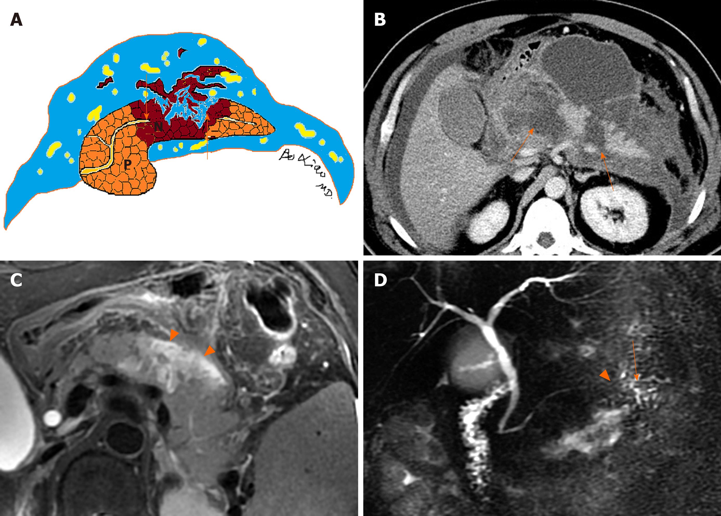
Figure 8 Schematic diagram of disconnected pancreatic duct syndrome and medical images of patients.
A: Schematic diagram of disconnected pancreatic duct syndrome. The pancreatic duct rupture and interruption (arrows) resulting from a wide range of liquefied pancreatic body tissue is seen; B: A 60-year-old man with necrotizing pancreatitis. Axial contrast-enhanced computed tomography image in the venous phase shows extensive parenchymal transmural necrosis (arrows) in the region of neck and body of the pancreas. Disconnected main pancreatic duct was proved at surgery; C: A 57-year-old woman with necrotizing pancreatitis. Magnetic resonance imaging axial T2WI image shows a majority of liquefied necroses (arrowheads) in the pancreatic body; D: The main pancreatic duct (arrow) of the tail of pancreas is interrupted (arrowhead) by the mentioned-above lesion. P: Pancreas; N: Necrosis.
Figure 9 Schematic diagram of necrotizing pancreatitis and computed tomography image of a patient.
A: Schematic diagram of necrotizing pancreatitis (peripancreatic necrotic type). Acute necrotic collection involving peripancreatic fat only (arrows) is seen; B: A 58-year-old woman with necrotizing pancreatitis (peripancreatic necrosis only). Axial contrast-enhanced computed tomography image in the venous phase shows normal pancreatic parenchymal enhancement without definite necrosis. A peripancreatic collection may be misdiagnosed as “acute peripancreatic fluid collection.” However, multiple heterogeneous, nonliquid adipose components (arrows) are revealed among the collection. For this reason, it should be considered as acute necrotic collection. P: Pancreas; APFC: Acute peripancreatic fluid collection; ANC: Acute necrotic collection.
Figure 10 Schematic diagram of necrotizing pancreatitis and magnetic resonance imaging of a patient.
A: Schematic diagram of necrotizing pancreatitis (pancreatic parenchymal necrosis alone). The peripancreatic collections are homogeneous; B: A 47-year-old man with necrotizing pancreatitis complicated with haemorrhage. Magnetic resonance imaging axial T1WI image shows peripancreatic homogeneous fluid with greater hyperintense signal. For this reason, it may be misdiagnosed as “acute peripancreatic fluid collection.” However, the necrosis and haemorrhage (arrow) of the body and tail of the pancreas can be indicated. Therefore, the collection should be diagnosed as acute necrotic collection. P: Pancreas, N: Necrosis, H: Haemorrhage; APFC: Acute peripancreatic fluid collection; ANC: Acute necrotic collection.
Figure 11 Schematic diagram of acute necrotizing pancreatitis, and extrapancreatic fluid and computed tomography of a patient.
A: Schematic diagram of acute necrotizing pancreatitis and extrapancreatic fluid with extension within the pancreatic parenchyma (arrow); B: A 56-year-old woman with acute necrotizing pancreatitis. Axial contrast-enhanced computed tomography image in the late arterial phase shows a peripancreatic homogeneous collection (arrowheads). It may be misinterpreted as “acute peripancreatic fluid collection.” However, note the peripancreatic fluid extends into the parenchyma of the head and neck of the pancreas (arrow). Therefore, it should be diagnosed as an “acute necrotic collection.” P: Pancreas; APFC: Acute peripancreatic fluid collection; ANC: Acute necrotic collection.
- Citation: Xiao B. Acute pancreatitis: A pictorial review of early pancreatic fluid collections. Artif Intell Med Imaging 2020; 1(1): 40-49
- URL: https://www.wjgnet.com/2644-3260/full/v1/i1/40.htm
- DOI: https://dx.doi.org/10.35711/aimi.v1.i1.40


