©The Author(s) 2021.
World J Meta-Anal. Aug 28, 2021; 9(4): 333-341
Published online Aug 28, 2021. doi: 10.13105/wjma.v9.i4.333
Published online Aug 28, 2021. doi: 10.13105/wjma.v9.i4.333
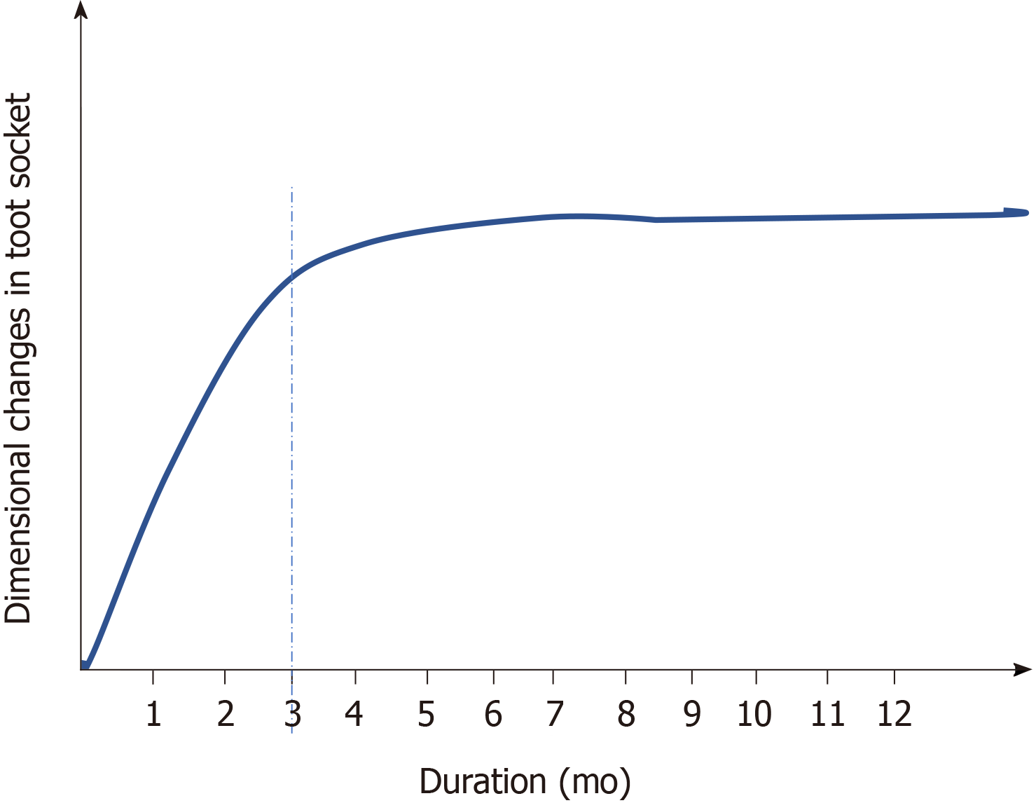
Figure 1 Dimensional changes as a part socket healing.
This graph shows that after tooth extraction greater degree of bone modelling occurs in first 3-mo and the remodeling continue later on for a year and beyond.
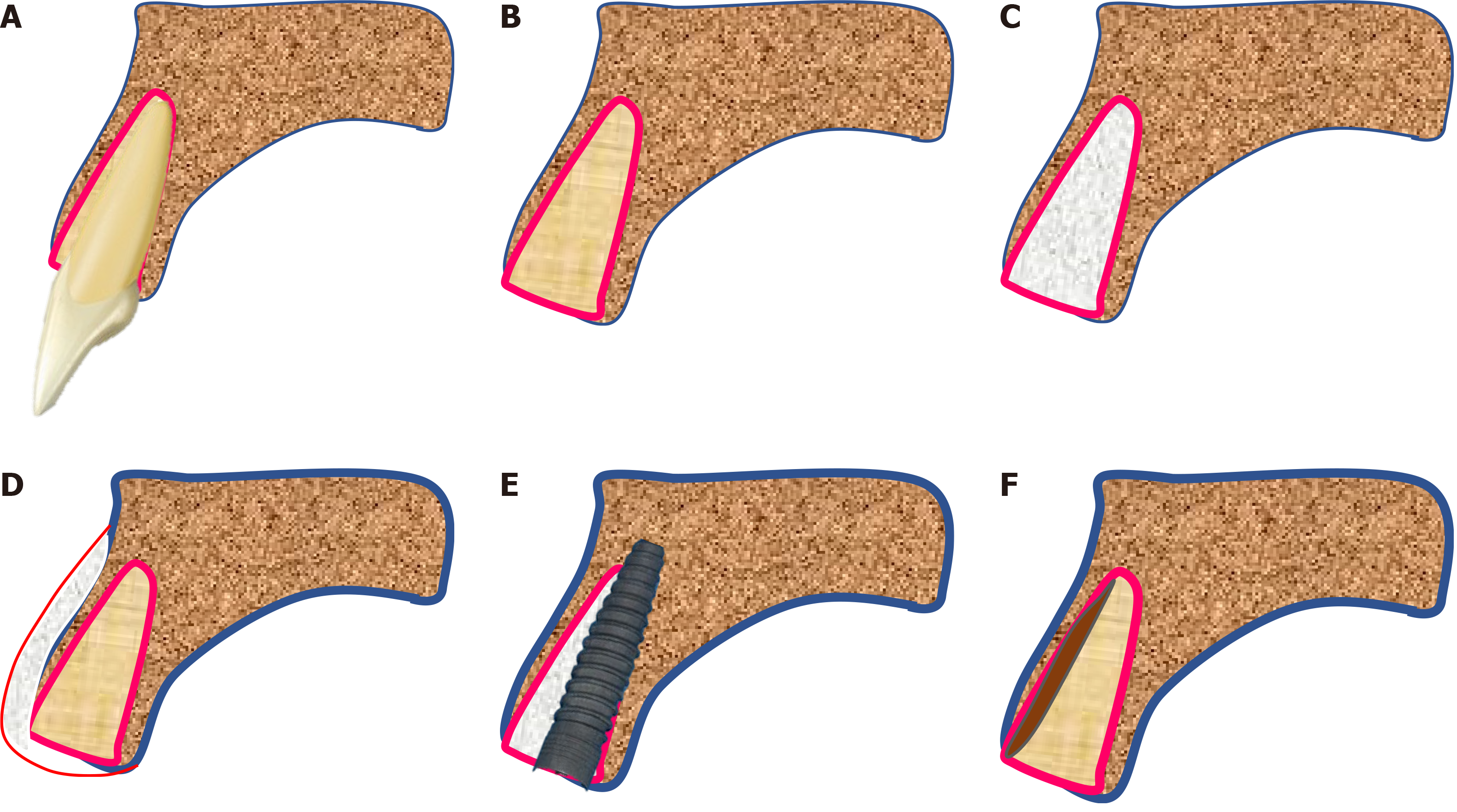
Figure 2 Techniques to compensate for the loss of socket wall post extraction.
A: Tooth in socket; B: Empty socket after the tooth is extracted; C: Socket grafting (with lots of different biomaterials) to preserve the alveolar bone; D: Buccal bone grafting with GTR (guided tissue regeneration) to increase the thickness of buccal bone; E: Placement of immediate implant (with/without grafting the jumping space); F: Retaining only a part of root in contact with the buccal bone plate (socket-shield).
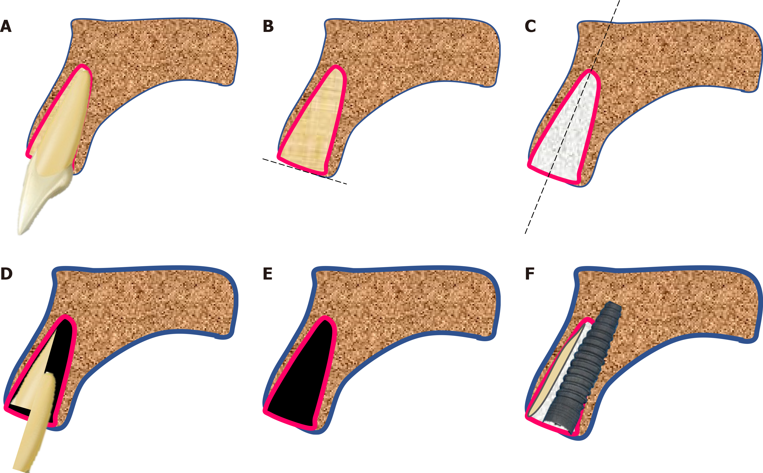
Figure 3 The ‘Socket-shield’ technique step-by-step.
A: Tooth in bony socket; B: The tooth in concern is sectioned horizontally 1 mm above the bone crest; C: Root is sectioned longitudinally in facial and palatal halves; D: The palatal section is extracted; E: The facial root section is concaved with a long shank dental bur; F: Immediate placement of dental implant palatal to the retained root fragment. The jumping space if any can be grafted is possible.
- Citation: Agrawal AA. Fate of root shell after pontic/socket shield techniques, is it better to extract the whole tooth? World J Meta-Anal 2021; 9(4): 333-341
- URL: https://www.wjgnet.com/2308-3840/full/v9/i4/333.htm
- DOI: https://dx.doi.org/10.13105/wjma.v9.i4.333


