©The Author(s) 2020.
World J Meta-Anal. Mar 28, 2020; 8(2): 109-118
Published online Mar 28, 2020. doi: 10.13105/wjma.v8.i2.109
Published online Mar 28, 2020. doi: 10.13105/wjma.v8.i2.109
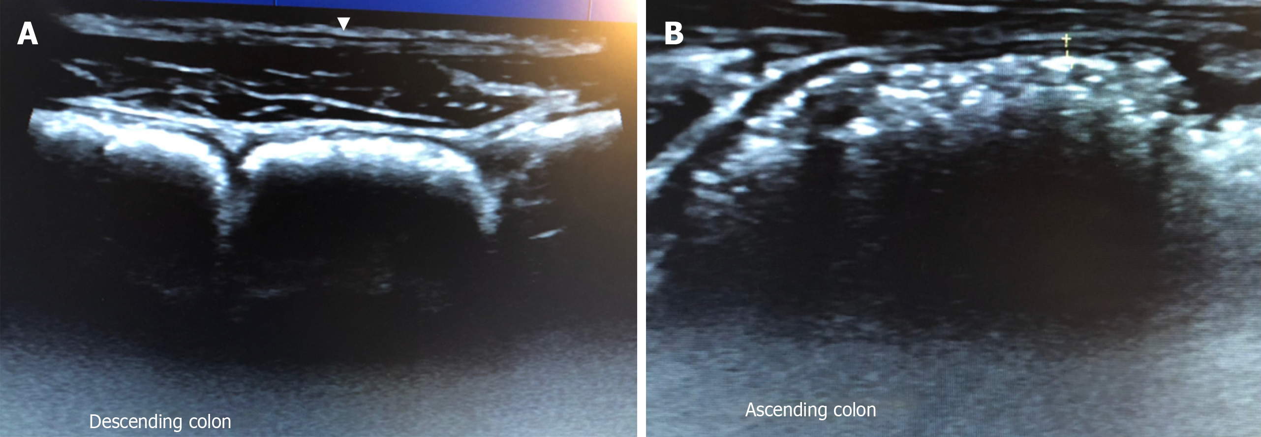
Figure 1 Gastrointestinal ultrasound images.
A: Gastrointestinal ultrasound images of descending colon showing haustra-shaped reflections with acoustic shadow behind, suggestive of severe faecal loading B: Gastrointestinal ultrasound images of strong reflection with acoustic shadow behind, also suggestive of faecal loading.
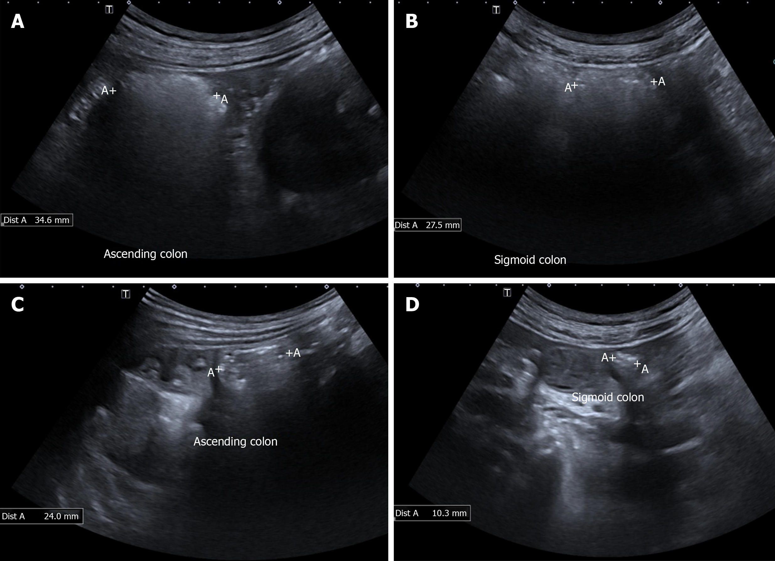
Figure 2 shows the ascending colon and sigmoid colon images of a constipated patients.
A: Image of ascending colon pre-treatment; B: Image of sigmoid colon pre-treatment; Note the strong reflections with acoustic shadow behind, suggestive of severe faecal loading; Colonic diameters are 34.6 mm and 27.5 mm respectively for ascending and sigmoid colon; C: Image of ascending colon post-treatment; D: Image of sigmoid colon post-treatment; Note the absence of strong reflections or acoustic shadows suggestive of minimal faecal loading; Colonic diameters of both ascending and sigmoid colon are also reduced post-treatment.
- Citation: Ong AML. Utility of gastrointestinal ultrasound in functional gastrointestinal disorders: A narrative review. World J Meta-Anal 2020; 8(2): 109-118
- URL: https://www.wjgnet.com/2308-3840/full/v8/i2/109.htm
- DOI: https://dx.doi.org/10.13105/wjma.v8.i2.109


