©The Author(s) 2019.
World J Meta-Anal. Feb 22, 2019; 7(2): 51-65
Published online Feb 22, 2019. doi: 10.13105/wjma.v7.i2.51
Published online Feb 22, 2019. doi: 10.13105/wjma.v7.i2.51
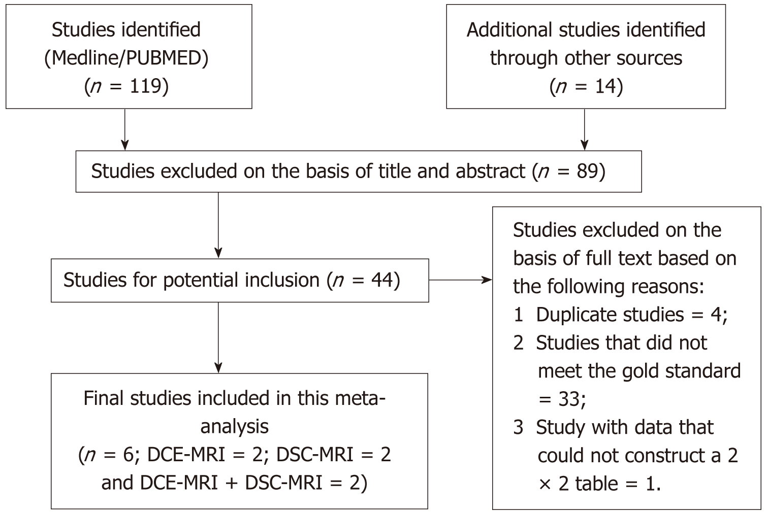
Figure 1 Flowchart outlining the study selection process according to the Preferred Reporting Items for Systematic Reviews guidelines.
DCE-MRI: Dynamic contrast-enhanced magnetic resonance imaging; DSC-MRI: Dynamic susceptibility contrast magnetic resonance imaging.
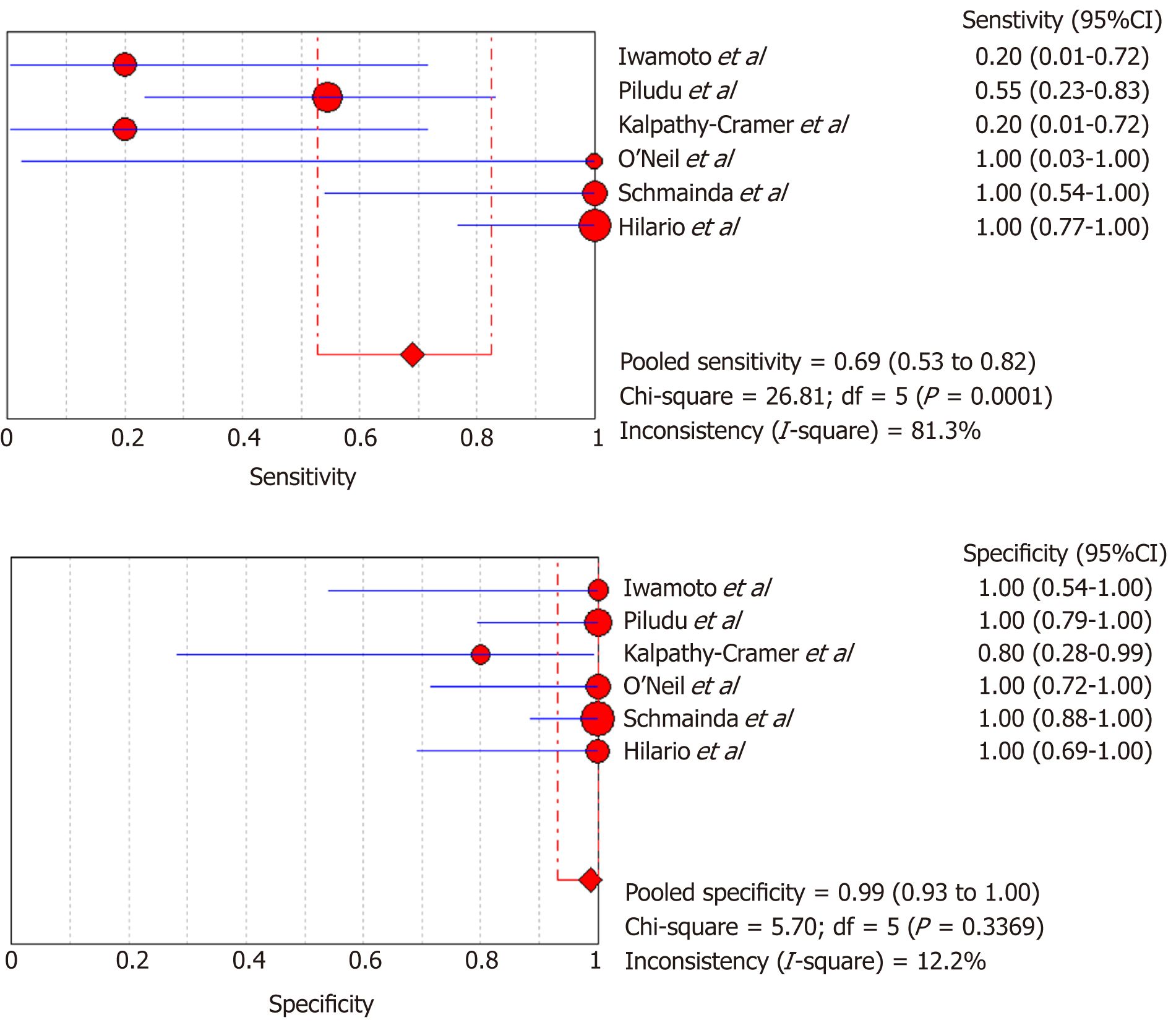
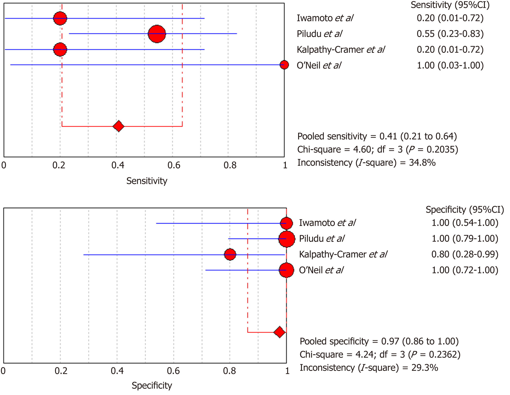
Figure 2 Sensitivity and specificity of dynamic contrast-enhanced magnetic resonance imaging and dynamic susceptibility contrast magnetic resonance imaging in the evaluation of response to antiangiogenic therapy in recurrent gliomas.
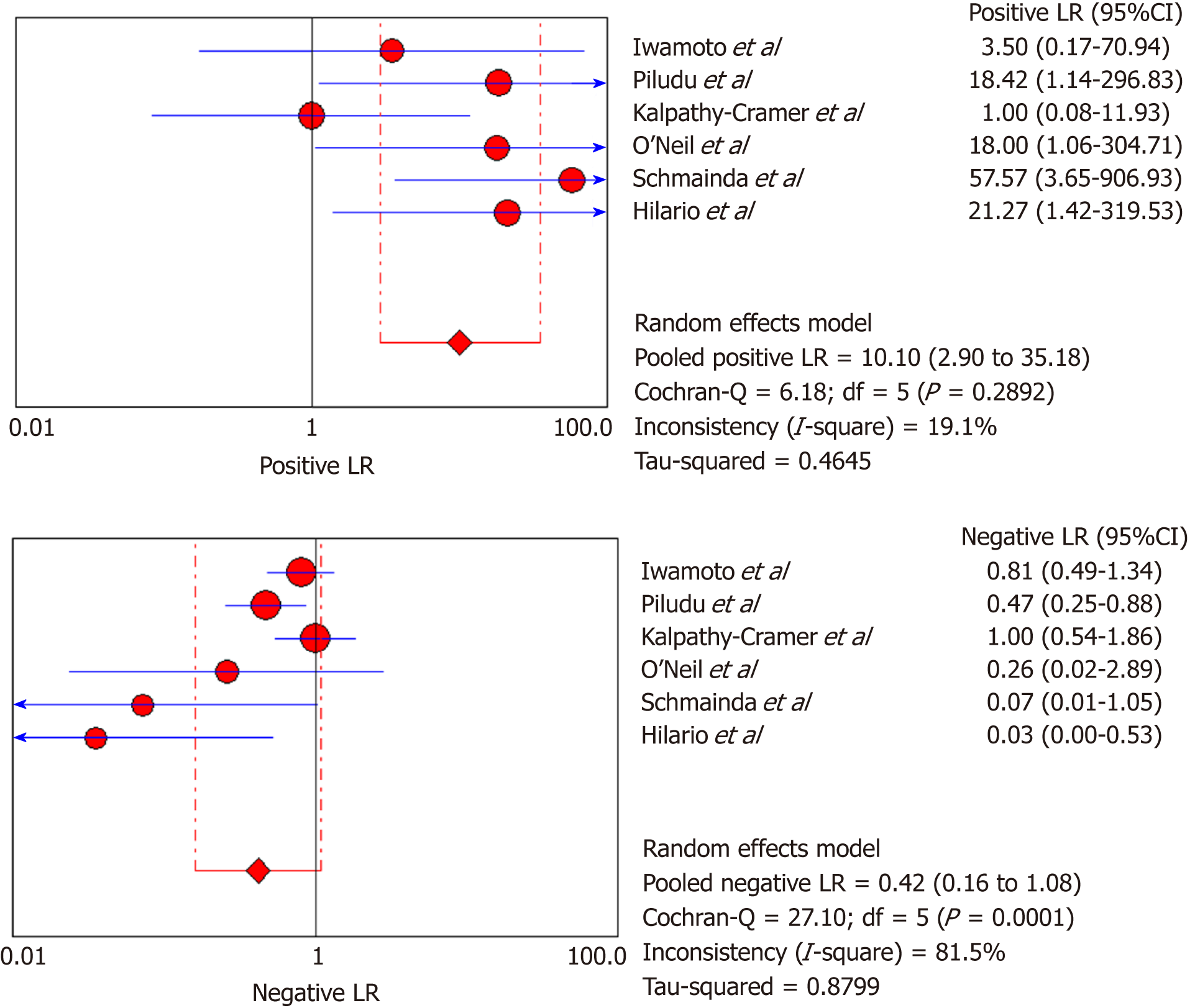
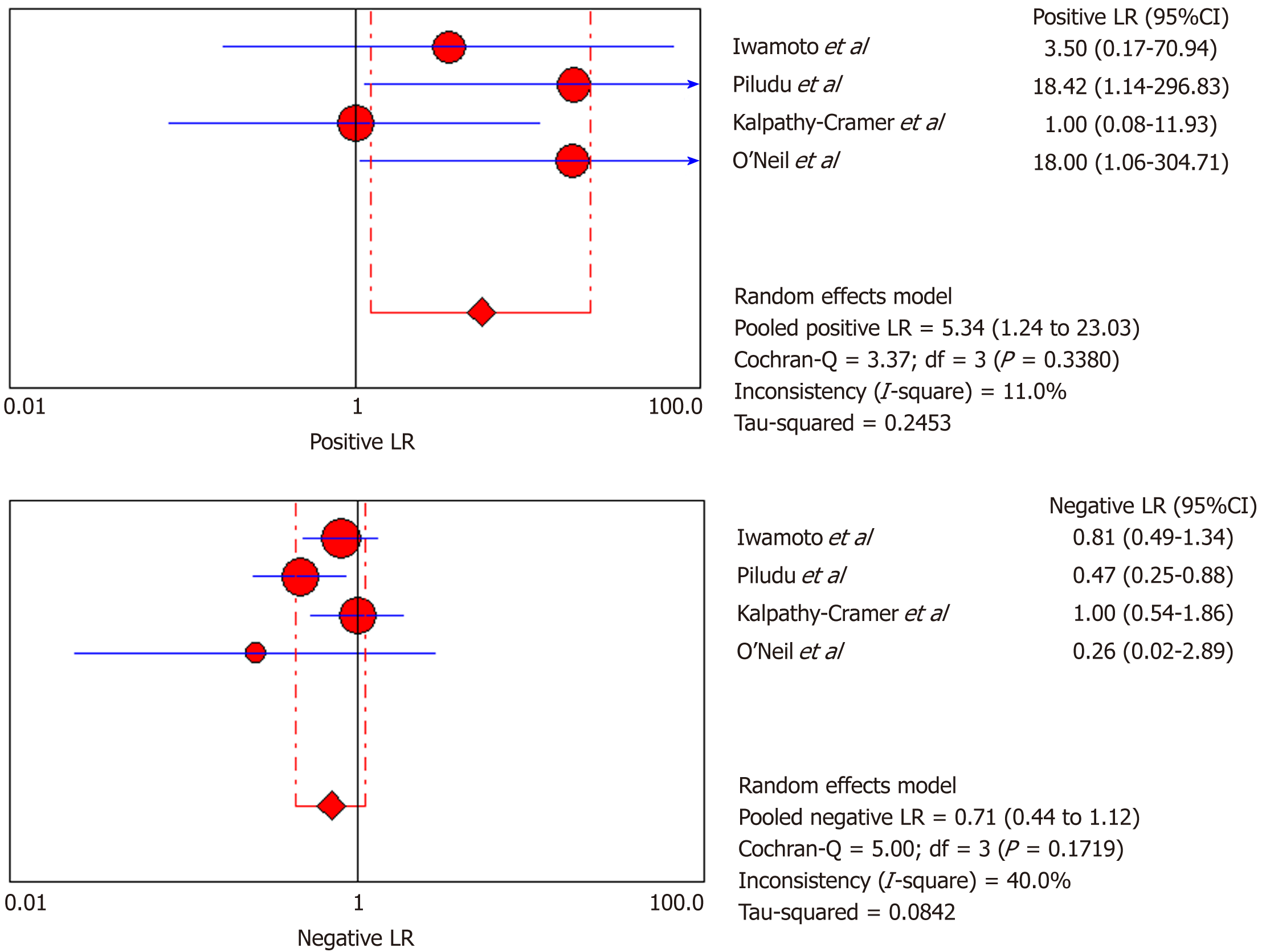
Figure 3 Likelihood ratio + and likelihood ratio - of dynamic contrast-enhanced magnetic resonance imaging and dynamic susceptibility contrast magnetic resonance imaging in the evaluation of response to antiangiogenic therapy in recurrent gliomas.
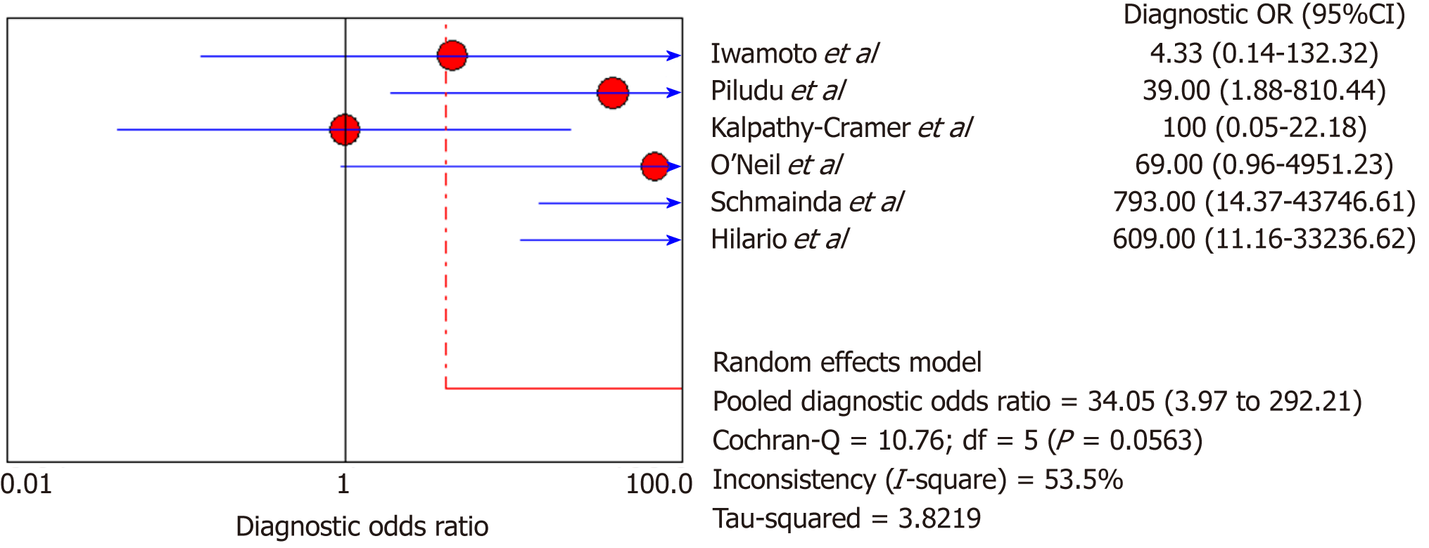
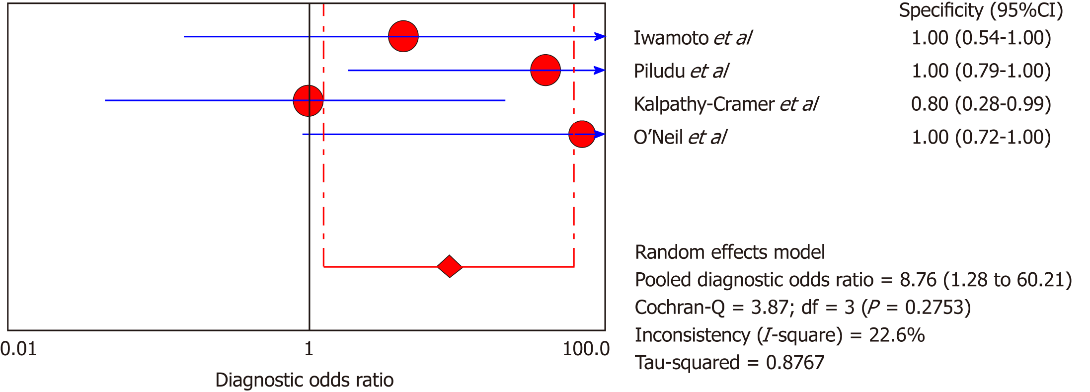
Figure 4 Diagnostic odds ratio of dynamic contrast-enhanced magnetic resonance imaging and dynamic susceptibility contrast magnetic resonance imaging in the evaluation of response to antiangiogenic therapy in recurrent gliomas.
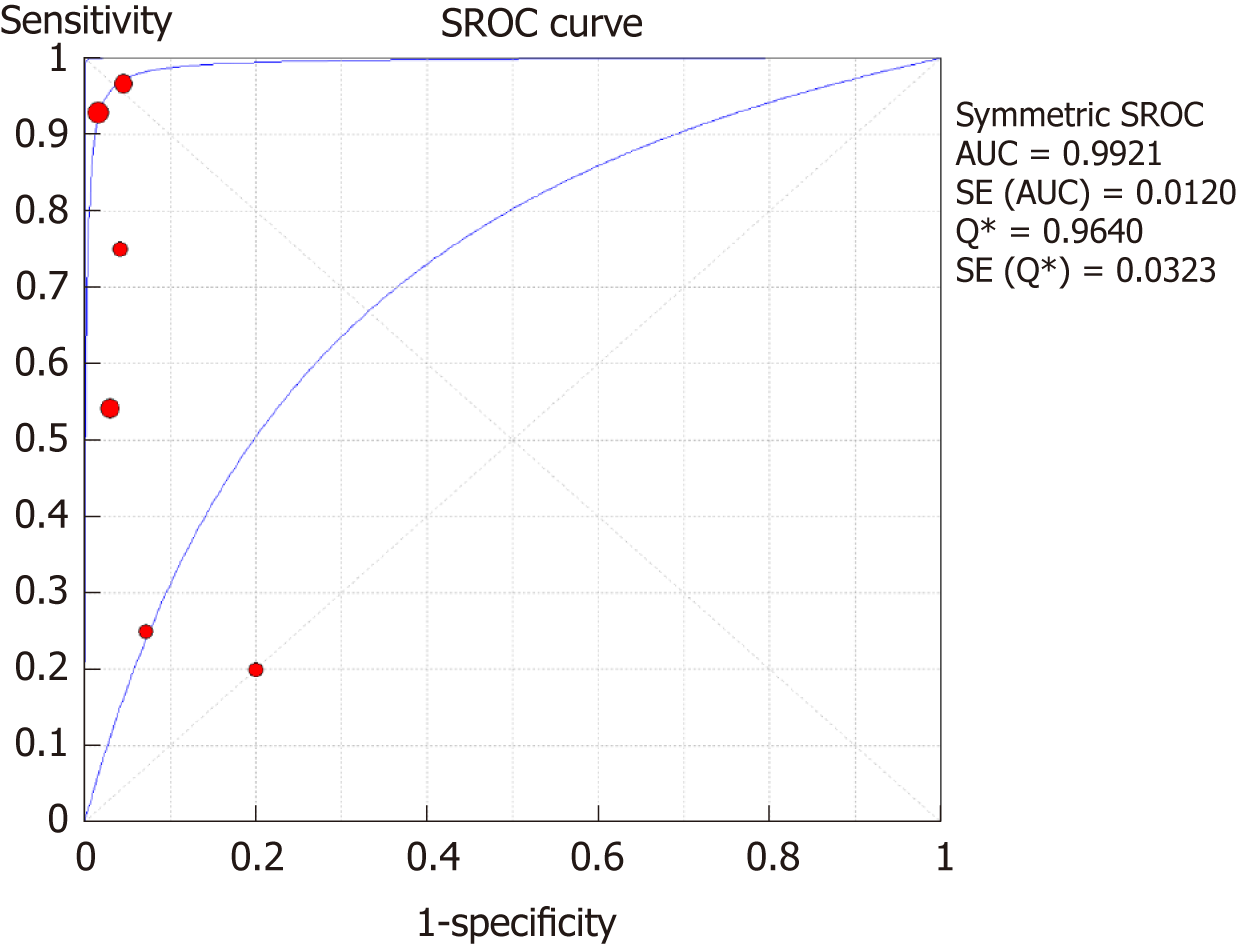
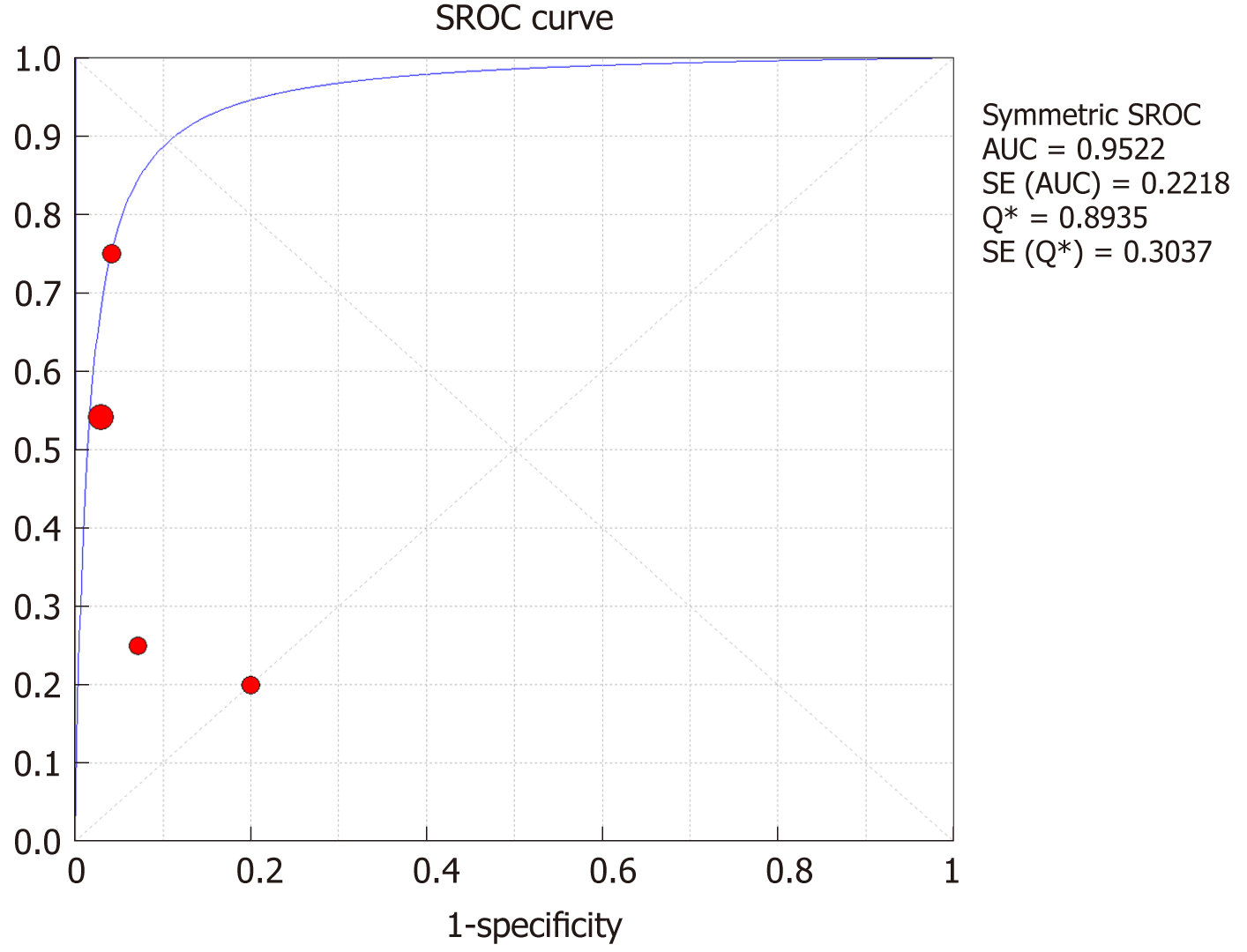
Figure 5 Summary receiver operating characteristic curve for dynamic contrast-enhanced magnetic resonance imaging and dynamic susceptibility contrast magnetic resonance imaging.
Solid circle represents each study in the meta-analysis, and the circle size indicates the study size. SROC: Summary receiver operating characteristic.
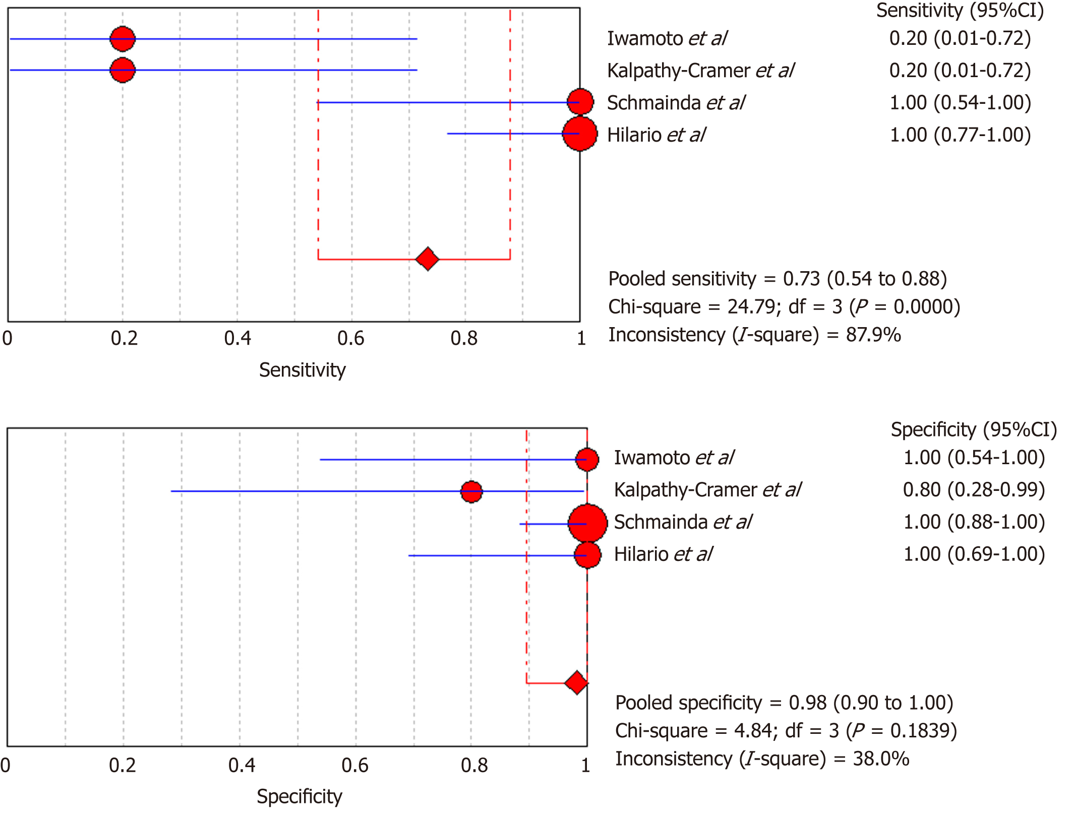
Figure 6 Sensitivity and specificity of dynamic susceptibility contrast magnetic resonance imaging in the evaluation of response to antiangiogenic therapy in recurrent gliomas.
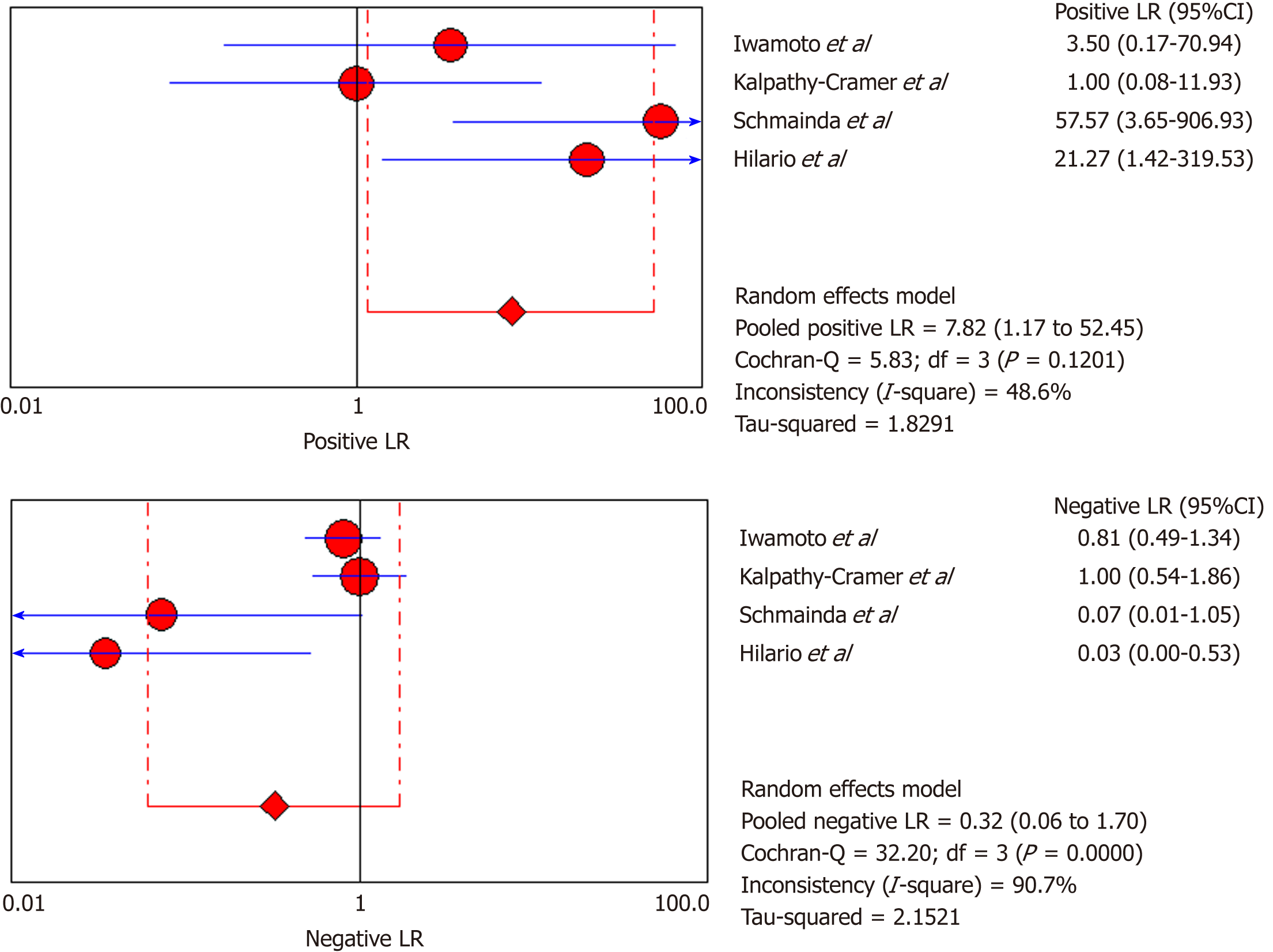
Figure 7 Likelihood ratio + and likelihood ratio - of dynamic susceptibility contrast magnetic resonance imaging in the evaluation of response to antiangiogenic therapy in recurrent gliomas.
Figure 8 Diagnostic odds ratio of dynamic susceptibility contrast magnetic resonance imaging in the evaluation of response to antiangiogenic therapy in recurrent gliomas.
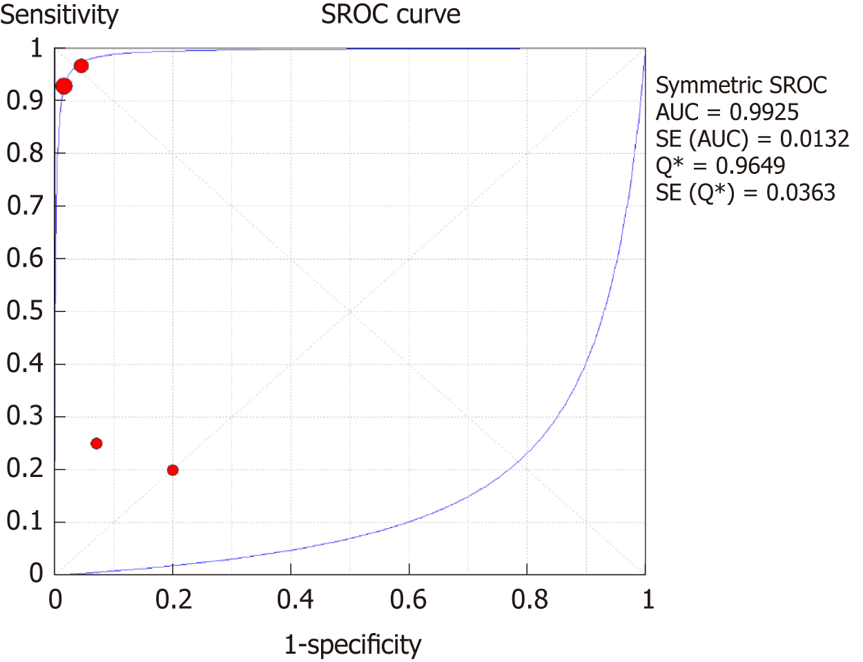
Figure 9 Summary receiver operating characteristic curve for dynamic susceptibility contrast magnetic resonance imaging.
SROC: Summary receiver operating characteristic.
Figure 10 Sensitivity and specificity of dynamic contrast-enhanced magnetic resonance imaging in the evaluation of response to antiangiogenic therapy in gliomas.
Figure 11 Likelihood ratio + and likelihood ratio - of dynamic contrast-enhanced magnetic resonance imaging in the evaluation of response to antiangiogenic therapy in recurrent gliomas.
Figure 12 Diagnostic odds ratio of dynamic contrast-enhanced magnetic resonance imaging in the evaluation of response to antiangiogenic therapy in recurrent gliomas.
Figure 13 Summary receiver operating characteristic curve for dynamic contrast-enhanced magnetic resonance imaging.
SROC: Summary receiver operating characteristic.
- Citation: Kasenene A, Baidya A, Shams S, Xu HB. Evaluation of tumor response to antiangiogenic therapy in patients with recurrent gliomas using contrast-enhanced perfusion-weighted magnetic resonance imaging techniques: A meta-analysis. World J Meta-Anal 2019; 7(2): 51-65
- URL: https://www.wjgnet.com/2308-3840/full/v7/i2/51.htm
- DOI: https://dx.doi.org/10.13105/wjma.v7.i2.51


