©The Author(s) 2023.
World J Clin Cases. Feb 16, 2023; 11(5): 1182-1187
Published online Feb 16, 2023. doi: 10.12998/wjcc.v11.i5.1182
Published online Feb 16, 2023. doi: 10.12998/wjcc.v11.i5.1182
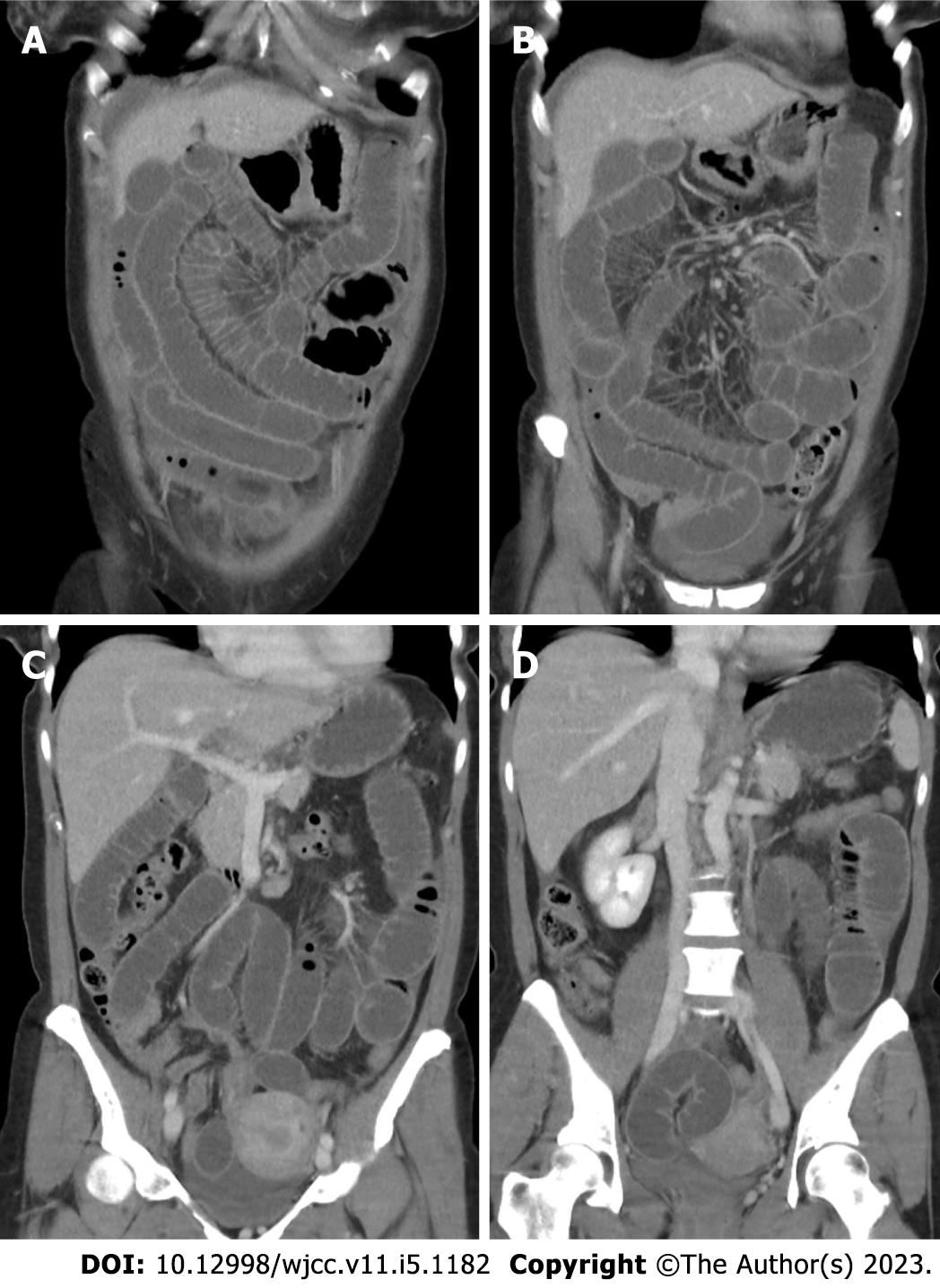
Figure 1 computer tomography scan of the abdomen.
A: Small bowel distension is observed; B: The dilated small bowel indicates an obstruction; C: The site of change of intestinal caliber is located in the right lower quadrant; D: The cause of obstruction cannot be identified.
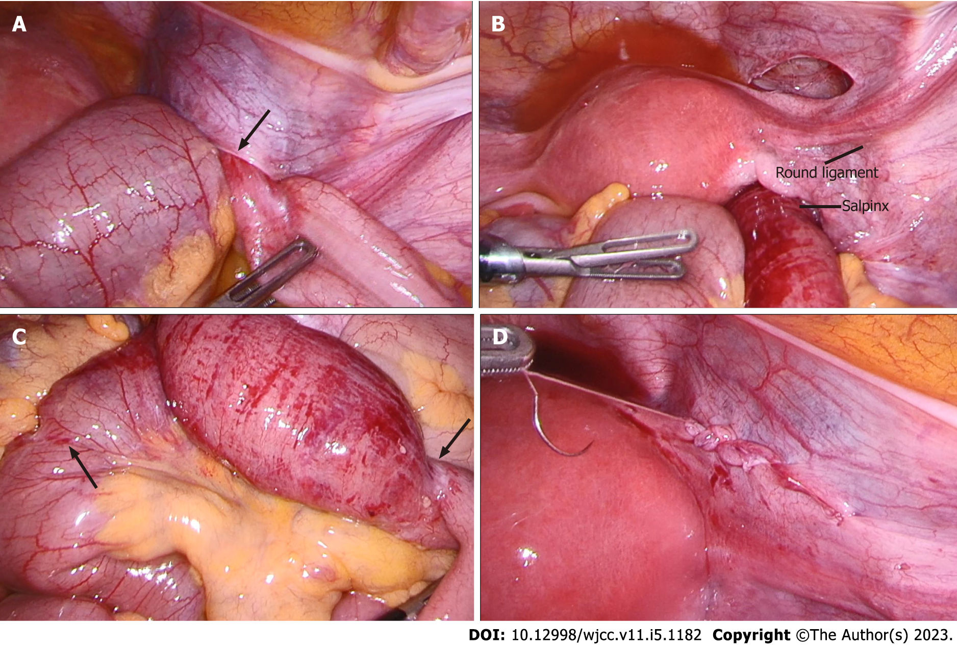
Figure 2 Intraoperative findings.
A: Internal hernia is identified: small bowel incarceration through a defect in the right broad ligament of the uterus (arrow); B: Overview on the anatomy: The defect is caudal to the salpinx and round ligament of the uterus. According to the classification by Cilley et al[13], this classifies as a type I hernia; C: Strangulation marks are indicated by arrows and between which hyperemic bowel can be observed; D: Defect closure with a running suture.
- Citation: Zucal I, Nebiker CA. Closed loop ileus caused by a defect in the broad ligament: A case report. World J Clin Cases 2023; 11(5): 1182-1187
- URL: https://www.wjgnet.com/2307-8960/full/v11/i5/1182.htm
- DOI: https://dx.doi.org/10.12998/wjcc.v11.i5.1182


