©The Author(s) 2023.
World J Clin Cases. Nov 6, 2023; 11(31): 7712-7717
Published online Nov 6, 2023. doi: 10.12998/wjcc.v11.i31.7712
Published online Nov 6, 2023. doi: 10.12998/wjcc.v11.i31.7712
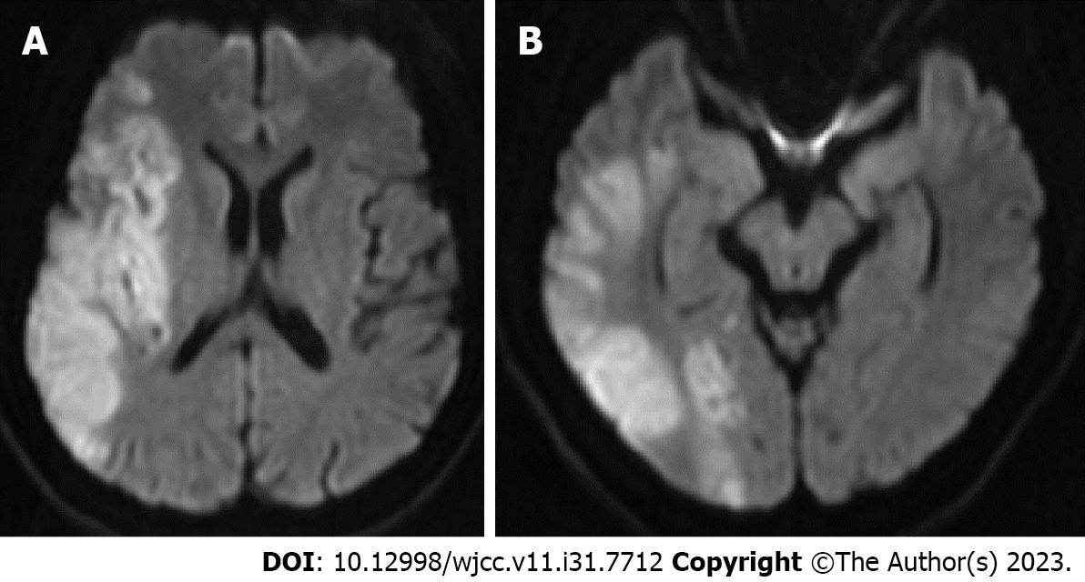
Figure 1 Diffusion-weighted imaging.
A: High signal intensity lesion in the territory of the right middle cerebral artery; B: High signal intensity lesion in some territory of the right posterior cerebral artery.
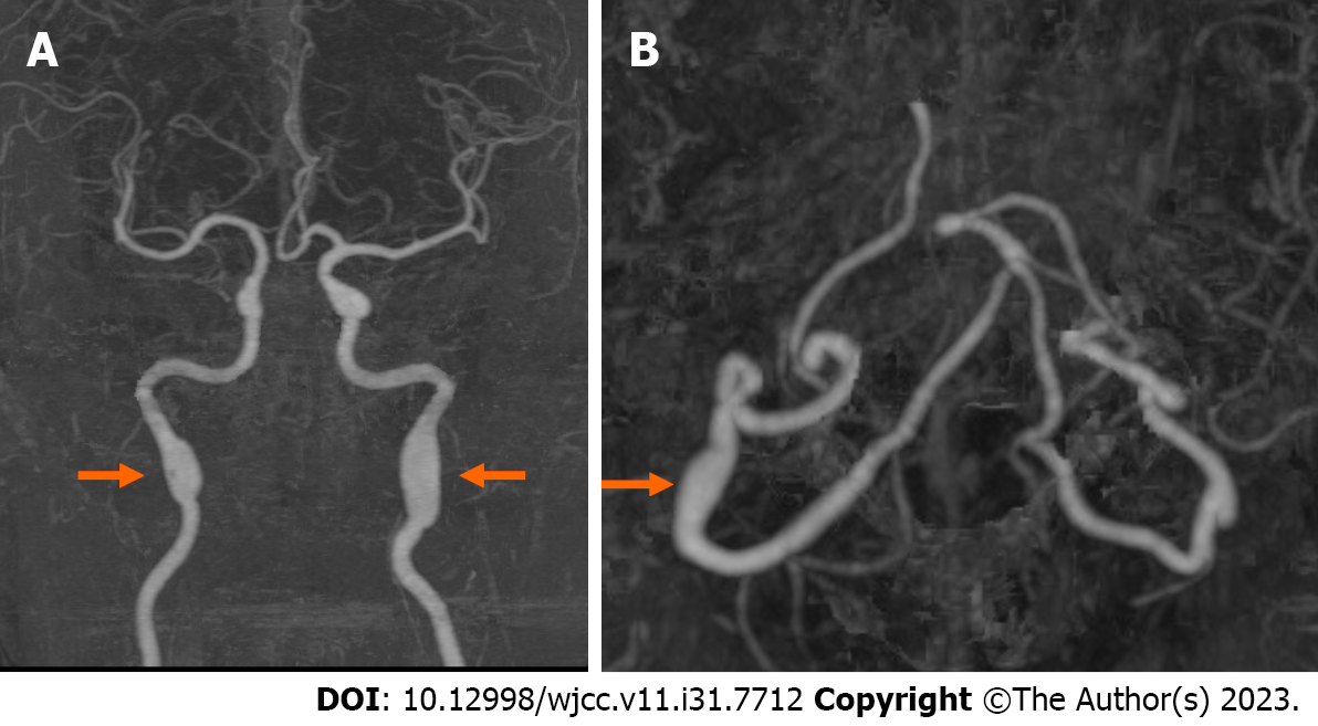
Figure 2 Computed tomography angiography.
A: Fusiform aneurysms in the petrous segment of bilateral internal carotid arteries (arrows); B: A Fusiform aneurysm in the V3 segment of the right distal vertebral artery (arrow).
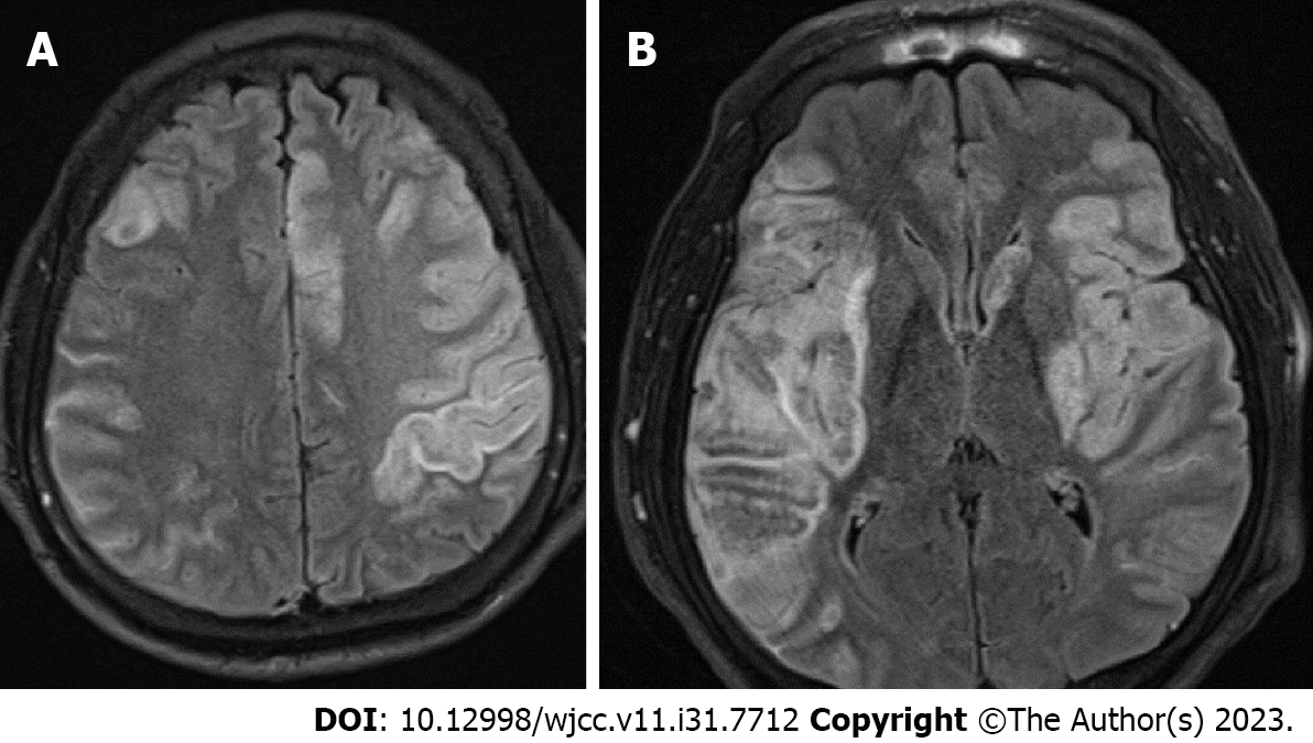
Figure 3 Magnetic resonance imaging.
A: New ischemic lesion in the left anterior cerebral artery; B: New ischemic lesion in the left middle cerebral artery with hemorrhagic transformed index lesion.
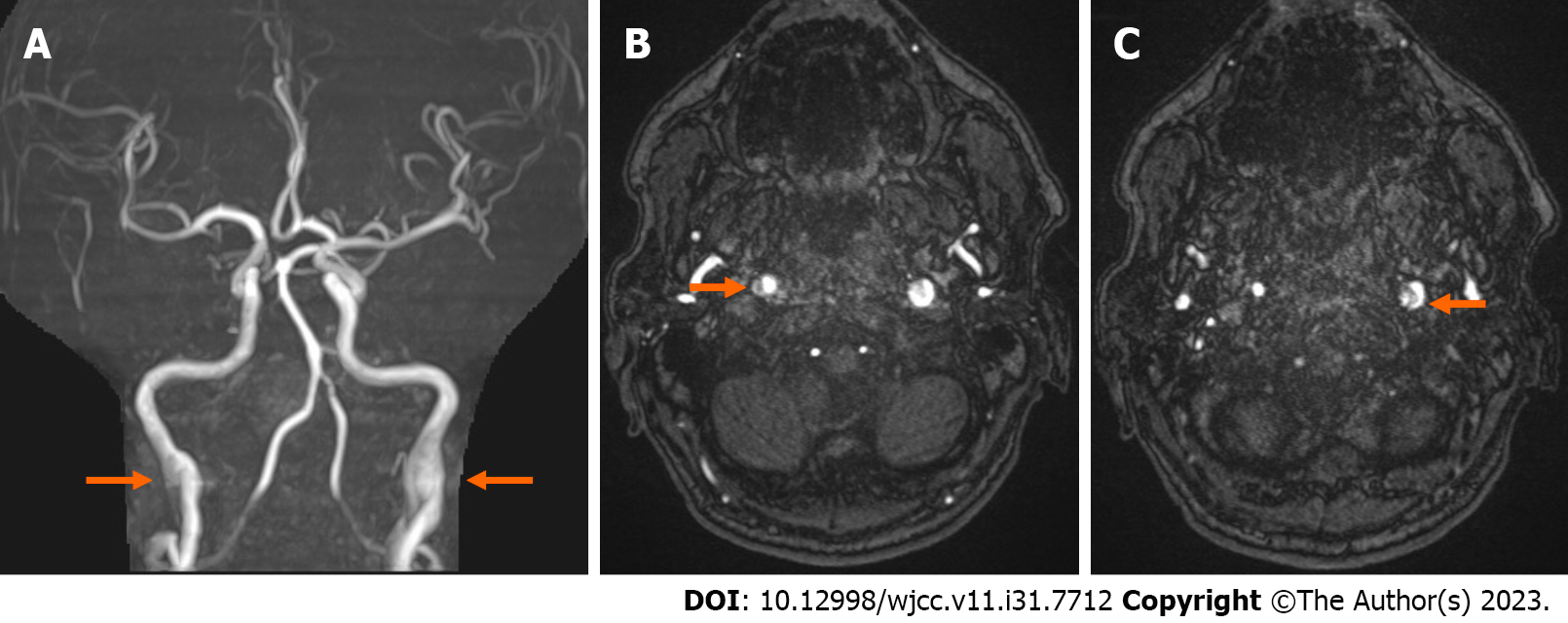
Figure 4 Magnetic resonance angiography.
A: Fusiform aneurysms of bilateral internal carotid arteries (ICAs; arrows); B and C: Double lumen appearance of bilateral ICAs (arrows).
- Citation: Shin DS, Yeo DK, Choi EJ. Successive development of ischemic malignant strokes in a patient with multiple fusiform aneurysms: A case report. World J Clin Cases 2023; 11(31): 7712-7717
- URL: https://www.wjgnet.com/2307-8960/full/v11/i31/7712.htm
- DOI: https://dx.doi.org/10.12998/wjcc.v11.i31.7712


