Published online Dec 26, 2017. doi: 10.5662/wjm.v7.i4.148
Peer-review started: January 18, 2017
First decision: March 27, 2017
Revised: November 18, 2017
Accepted: December 1, 2017
Article in press: December 1, 2017
Published online: December 26, 2017
Processing time: 343 Days and 13.6 Hours
The potential complications associated with an adnexal mass discovered during early pregnancy call for surgical treatment. Ideally, surgery is performed after gestational week 12, but uterine expansion after the first trimester makes surgery difficult. We report two pregnancies complicated by adnexal masses for which we used an organ fixation device for safe performance of single-site umbilical laparoscopic surgery. Pelvic magnetic resonance imaging depicted a dichorionic, diamniotic twin pregnancy and 60-mm right adnexal mass in the first patient and bilateral adnexae in the second. All three masses were suspected mature cystic teratomas. Both patients underwent laparoscopic surgery during gestational week 14. With use of an organ fixation device, traction was applied until the mass reached the umbilicus; tumor resection was performed extracorporeally. In the second patient, the second mass was simply aspirated because adhesions were encountered. Our single-site laparoscopic-extracorporeal technique proved to be a safe approach to an otherwise high-risk situation.
Core tip: The new device, “Ova-Lead” has a 20-mm-diameter tip that is made of silicone and shaped like a suction cup. It fixes to the organ through the application of negative pressure, by using this device the surgeon manipulates the organ. We reported two cases of adnexal mass discovered during pregnant that this device seemed useful.
- Citation: Kasahara H, Kikuchi I, Otsuka A, Tsuzuki Y, Nojima M, Yoshida K. Laparoscopic-extracorporeal surgery performed with a fixation device for adnexal masses complicating pregnancy: Report of two cases. World J Methodol 2017; 7(4): 148-150
- URL: https://www.wjgnet.com/2222-0682/full/v7/i4/148.htm
- DOI: https://dx.doi.org/10.5662/wjm.v7.i4.148
One or more adnexal masses are discovered in a reported 0.01%-1% of all pregnancies[1]. The majority resolve spontaneously, but in some cases, torsion or rupture necessitates emergency surgery. Ideally, surgery is performed after gestational week 12, but expansion of the uterus after the first trimester makes surgical manipulation difficult. We report adnexal masses complicating 2 pregnancies for which we used an organ fixation device for safe performance of single-site umbilical laparoscopic surgery.
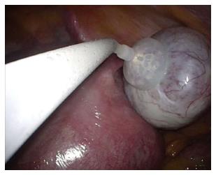
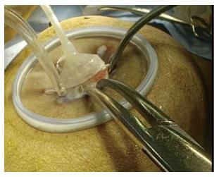
The patient was a 29-year-old woman, gravida 0 para 0. An early-stage dichorionic, diamniotic twin pregnancy was confirmed simultaneously with a right-sided adnexal mass. Pelvic magnetic resonance imaging (MRI) revealed a 60-mm mass that appeared to be a mature cystic teratoma, so laparoscopic surgery was scheduled and performed during gestational week 14. We placed Lap Protector and EZ Access (Hakko Corporation Tokyo Japan) as a wound retractor in the umbilicus, and we used an organ fixation device called “Ova-Lead” (Fuji Systems corporation, Tokyo Japan) to apply traction to the adnexa to bring the mass up to the umbilicus (Figure 1). We resected the mass extracorporeally (Figure 2). She was followed up at our hospital until 22 wk then moved overseas.
The patient was a 38-year-old woman, gravida 0 para 0. Bilateral adnexal masses were identified during the early stage of pregnancy. Pelvic MRI revealed a 70-mm left adnexal mass and a 50-mm right adnexal mass. Both were thought to be mature cystic teratomas, so laparoscopic surgery was scheduled and performed during gestational week 14. We placed a RapidPort EZ Access Port in the umbilicus. We encountered extensive adhesions within the peritoneal cavity. Traction was applied to the left adnexa by means of an organ fixation device, “Ova-Lead” until the mass reached the umbilicus, and the mass was resected extracorporeally. Applying traction to the right adnexa proved difficult due to the adhesions, so we simply performed paracentesis. The fluid contained hemorrhagic components, so we suspected an endometrioma. The left adnexal mass was diagnosed histologically as a mixed cystic teratoma and endometrioma. No recurrence of the right mass was noted during the remaining course of the pregnancy. She was followed up at our hospital, and at 36 wk, premature rupture of membrane occurred to her, then she was delivered vaginally.
The “Ova-Lead” has a 20-mm-diameter tip that is made of silicone and shaped like a suction cup. It fixes to the organ through the application of negative pressure (30 mmHg). The surgeon manipulates the device by a metal handle, and use of the device eases performance of the operation. At our hospital, we attach and fix the device to the adnexal mass and then apply traction to the mass to draw it up to the umbilicus. This allows us to resect the organ extracorporeally, and by using this single-port technique, the operation time is shortened, and surgery can be performed with as little leakage of tumor contents into the peritoneal cavity as possible.
Adnexal masses occurring during pregnancy are of various tissue types. The most common are mature cystic teratomas, representing 40% of adnexal masses. These are followed in order by serous cystadenomas at 20%, mucinous cystadenomas at 10%, endometriomas at 5%, and malignant tumors at 3%[1]. The contents of a mature cystic teratoma can easily leak into the peritoneal cavity. This has been found to result in chemical peritonitis. Therefore, at our hospital, we use an extracorporeal technique to resect the mass.
Extracorporeal resection generally reduces the overall operation time, thus shortening the pneumoperitoneum time. Jansen et al[2] verified that increases in intrauterine pressure can result in fetal hypoxia. Hunter et al[3] verified in an animal model that carbon dioxide (gas) pneumoperitoneum can cause fetal acidosis and noted that changes were greatest at pressures of 15 mmHg or more. We believe that laparoscopic-extracorporeal resection, in comparison to total intracorporeal laparoscopic resection, allows us to better shorten the duration of both the surgery and the pneumoperitoneum, helping us prevent this complication.
Adverse events can occur in pregnant women with an adnexal mass. There is potential for pedicle torsion (1%-22%), rupture (9%), miscarriage or premature labor (5%-15%), or infection (1.2%-2.4%)[4]. The risk of such complications increases when the mass is greater than 6 cm[4]. We recommend surgery for masses greater than 6 cm that we encounter at our hospital.
Our gynecology department guidelines recommend performing surgery after gestational week 12, after the period of organogenesis has passed. The Japan College of Radiology imaging guidelines recommend use of MRI after gestational week 14. Surgery becomes increasingly difficult with each passing gestational week, so at our hospital, we perform surgical treatment as close to gestational week 14 as possible if a diagnosis has been made by then.
In conclusion, we treated two cases of adnexal masses complicating pregnancy by performing single-port umbilical laparoscopic surgery, using an organ fixation device, and resecting the masses extracorporeally. There are risks associated with surgery performed during pregnancy, but the potential complications associated with the simultaneous presence of an adnexal mass outweigh the risks of surgery. We have found that our technique prevents the complications associated with such surgery and facilitates safe surgical treatment even when the surgical field is difficult to secure and the uterus is especially enlarged, as in the case of a twin pregnancy. And also, even in the case of a large ovarian tumor, this method was suggested to be useful.
Ovarian cyst (benign).
Ovarian carcinoma, etc.
Magnetic resonance imaging findings as follows: Case 1: Mature cystic teratoma; Case 2: Endometrioma.
Case 1: Mature cystic teratoma; Case 2: Endometrioma.
Surgical treatment.
This manuscript was the first report about this device.
In the case of a large ovarian tumor, this “ova-lead” was suggested to be useful.
| 1. | Hoover K, Jenkins TR. Evaluation and management of adnexal mass in pregnancy. Am J Obstet Gynecol. 2011;205:97-102. [RCA] [PubMed] [DOI] [Full Text] [Cited by in Crossref: 67] [Cited by in RCA: 64] [Article Influence: 4.3] [Reference Citation Analysis (0)] |
| 2. | Jansen CA, Krane EJ, Thomas AL, Beck NF, Lowe KC, Joyce P, Parr M, Nathanielsz PW. Continuous variability of fetal PO2 in the chronically catheterized fetal sheep. Am J Obstet Gynecol. 1979;134:776-783. [RCA] [PubMed] [DOI] [Full Text] [Cited by in Crossref: 79] [Cited by in RCA: 76] [Article Influence: 1.6] [Reference Citation Analysis (0)] |
| 3. | Hunter JG, Swanstrom L, Thornburg K. Carbon dioxide pneumoperitoneum induces fetal acidosis in a pregnant ewe model. Surg Endosc. 1995;9:272-277; discussion 277-279. [RCA] [PubMed] [DOI] [Full Text] [Cited by in Crossref: 87] [Cited by in RCA: 74] [Article Influence: 2.4] [Reference Citation Analysis (0)] |
| 4. | Guariglia L, Conte M, Are P, Rosati P. Ultrasound-guided fine needle aspiration of ovarian cysts during pregnancy. Eur J Obstet Gynecol Reprod Biol. 1999;82:5-9. [RCA] [PubMed] [DOI] [Full Text] [Cited by in Crossref: 27] [Cited by in RCA: 28] [Article Influence: 1.0] [Reference Citation Analysis (0)] |
Open-Access: This article is an open-access article which was selected by an in-house editor and fully peer-reviewed by external reviewers. It is distributed in accordance with the Creative Commons Attribution Non Commercial (CC BY-NC 4.0) license, which permits others to distribute, remix, adapt, build upon this work non-commercially, and license their derivative works on different terms, provided the original work is properly cited and the use is non-commercial. See: http://creativecommons.org/licenses/by-nc/4.0/
Manuscript source: Invited manuscript
Specialty type: Medical laboratory technology
Country of origin: Japan
Peer-review report classification
Grade A (Excellent): 0
Grade B (Very good): B
Grade C (Good): C
Grade D (Fair): 0
Grade E (Poor): 0
P- Reviewer: Tu H, Zafrakas M S- Editor: Ji FF L- Editor: A E- Editor: Lu YJ


