Published online May 20, 2021. doi: 10.5662/wjm.v11.i3.88
Peer-review started: October 13, 2020
First decision: December 21, 2020
Revised: January 2, 2021
Accepted: March 11, 2021
Article in press: March 11, 2021
Published online: May 20, 2021
Processing time: 210 Days and 14 Hours
Mandibular fractures constitute about 80.79% of maxillofacial injuries in Alexandria University, either as isolated mandibular fractures or as a part of panfacial fractures. The combination of symphyseal and parasymphyseal frac
To compare the effectiveness of lag screws vs double Y-shaped miniplates in the fixation of anterior mandibular fractures.
This study is a prospective randomized controlled clinical trial, performed on sixteen patients with anterior mandibular fractures. Patients were divided equally into two groups, each consisting of eight patients. Group 1: Underwent open reduction and internal fixation using two lag screws. Group 2: Underwent open reduction and internal fixation using double Y-shaped plates. The following parameters were assessed: operating time in minutes, pain using a visual analog scale, edema, surgical wound healing for signs and symptoms of infection, occlusion status and stability, maximal mouth opening, and sensory nerve function. Cone beam computed tomography was performed at 3 and 6 mo to measure bone density and assess the progression of fracture healing.
The study included 13 males (81.3%) and 3 females (18.8%) aged 26 to 45 years (mean age was 35.69 ± 6.01 years). The cause of trauma was road traffic accidents in 10 patients (62.5%), interpersonal violence in 3 patients (18.8%) and other causes in 3 patients (18.8%). The fractures comprised 10 parasymphyseal fractures (62.5%) and 6 symphyseal fractures (37.5%). The values of all parameters were comparable in both groups with no statistically significant difference except for the mean bone density at 3 mo postoperatively which was 946.38 ± 66.29 in group 1 and 830.36 ± 95.53 in group 2 (P = 0.015).
Both lag screws and double Y-shaped miniplates provide favorable means of fixation for mandibular fractures in the anterior region. Fractures fixed with lag screws show greater mean bone density at 3 mo post-operation, indicative of higher primary stability and faster early bone healing. Further studies with larger sample sizes are required to verify these conclusions.
Core Tip: The aim of this study is to compare the effectiveness of lag screws vs double Y-shaped miniplates in the fixation of anterior mandibular fractures in terms of fracture stability and progression of bone healing.
- Citation: Melek L. Comparison of lag screws and double Y-shaped miniplates in the fixation of anterior mandibular fractures. World J Methodol 2021; 11(3): 88-94
- URL: https://www.wjgnet.com/2222-0682/full/v11/i3/88.htm
- DOI: https://dx.doi.org/10.5662/wjm.v11.i3.88
Mandibular fractures constitute about 80.79% of maxillofacial injuries in Alexandria University, either as isolated mandibular fractures or as a part of panfacial fractures. The combination of symphyseal and parasymphyseal fractures represent 47.09% of the total mandibular fractures[1]. However, this percentage of anterior mandibular frac
Lag screws have been described as a reliable, stable and safe method of internal fixation for anterior mandibular fractures. The absence of anatomical hazards, thickness of the bone cortices and curvature of the anterior mandible are all factors contributing to the suitability and success of using lag screws in that region[3].
Miniplates have been widely used for decades for the fixation of mandibular fractures owing to their easy handling and adaptation, in addition to providing functionally stable fixation[4]. Different designs of miniplates, varying from the conventional form by Champy, have been proposed to provide extra stability of the fracture. A biomechanical study has shown that double Y-shaped miniplates provide greater resistance to displacement in comparison to conventional straight miniplates[5].
The aim of this study is to compare the effectiveness of lag screws vs double Y-shaped miniplates in the fixation of anterior mandibular fractures.
This study is a prospective randomized controlled clinical trial. It was performed on sixteen patients with anterior mandibular fractures, selected from those admitted to the Emergency Department of Alexandria University Hospital. This study followed the Declaration of Helsinki with regard to medical protocol and ethics, and the regional Ethical Review Board of the Faculty of Dentistry, Alexandria University approved the study (Approval Number: IRB 00010556-IORG 0008839). The study was registered on clinicaltrials.gov (ClinicalTrials.gov ID: NCT04396054). A written informed consent was signed by each patient before the operation.
The patients were divided equally into two groups, each consisting of eight patients. Assignment of each patient into one of these two groups was carried out using computer random numbers: Group 1: Underwent open reduction and internal fixation using two lag screws; Group 2: Underwent open reduction and internal fixation using double Y-shaped plates.
Patients of both genders aged from 25 to 45 years, suffering from anterior fractures of the mandible (symphyseal or parasymphyseal) were included. Those with old frac
A thorough clinical examination was performed preoperatively on all patients, in addition to panoramic radiographs. All patients were operated by the same surgeon under general anaesthesia with nasotracheal intubation. Complete disinfection of the oral cavity and face was performed using povidone iodine solution, followed by draping with sterile towels exposing the surgical site. Maxillomandibular fixation was carried out to adjust the occlusion using arch bars and eyelet wiring. After that, an intraoral mandibular vestibular incision was made exposing the fracture line where reduction of the two segments was carried out under direct vision.
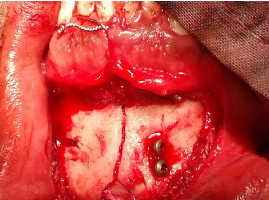
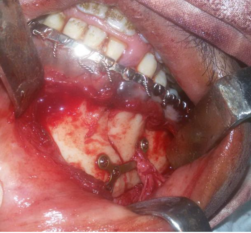
In the first group, fixation of the reduced segments was achieved using 2 lag screws (O and M Medical GmbH Eschenweg, Germany). The diameter of the screws was 2.7 mm and the length ranged from 18 to 24 mm. Screw fixation was performed by passage of the screw through a larger gliding hole into a smaller traction hole on each side of the fracture (Figure 1). In the second group, fixation of the reduced segments was achieved using double Y-shaped plates (Stryker-Leibenger, Germany) with 6 monocortical 2.0 mm diameter screws (Figure 2).
After direct fixation was performed in both groups, the incision was closed using layered suturing and the maxillomandibular fixation was removed. Postoperative care for all patients included the following: (1) Each patient received intravenous Cefotaxime 1 mg/12 h (Cefotax, by EIPICO) for one day postoperatively followed by Amoxicillin clavulanate (Augmentin, manufactured by MPU) 1 mg given orally twice daily for the next 5 d; (2) An analgesic anti-inflammatory drug in the form of Diclofenac Sodium (Rheumafen, by GlaxoSmithKline) 75 mg vial up to the second postoperative day was given followed by Diclofenac Potassium (Rheumafen tablets, by GlaxoSmithKline) 50 mg tablets three times daily for the next 5 d; (3) All patients were instructed to use chlorohexidine mouth wash (Hexitol, by an Arabic drug company) for maintenance of good oral hygiene; and (4) Instructions for a soft high calorie diet was given to all patients to be followed for 4 wk postoperatively.
Postoperative follow-up: Patients were followed up on the second, third post
Data were fed to the computer and analyzed by the appropriate statistical tests using the IBM Statistical Package for Social Science software version 21.0. Significance of the obtained results was set at the 5% level. Qualitative data were described using number and percent. Quantitative data were described using range (minimum and maximum), mean and standard deviation. The independent samples t-test was used to compare the means of quantitative data.
This study was conducted on 16 patients suffering from anterior mandibular fractures. The study included 13 males (81.3%) and 3 females (18.8%) aged 26 to 45 years (mean age was 35.69 ± 6.01 years). The cause of trauma was road traffic accidents in 10 patients (62.5%), interpersonal violence in 3 patients (18.8%) and other causes in 3 patients (18.8%). The fractures comprised 10 parasymphyseal fractures (62.5%) and 6 symphyseal fractures (37.5%).
In group 1, patients were treated with open reduction and internal fixation using lag screws and the mean operating time from start of hardware application to end of fixation was 14.38 ± 1.92 min. In group 2, patients were treated with open reduction and internal fixation using double Y-shaped miniplates and the mean operating time was 15.63 ± 1.53 min. The difference between the two groups regarding the mean operating time was statistically insignificant (P > 0.05) (Table 1).
| Group | n | mean ± SD | t | Significance (2-tailed) | |
| Operating time | 1 | 8 | 14.3750 ± 1.92261 min | 0.172 | |
| Operating time | 2 | 8 | 15.6250 ± 1.52947 min | -1.439 |
With regard to postoperative edema, only 2 patients in the study sample showed severe edema (12.5%), while all other patients demonstrated mild to moderate edema (87.5%) on the second postoperative day. By the end of the first week, the edema has resolved completely in all patients.
The mean pain intensity in the first postoperative week was 4.125 ± 1.25 in group 1 and 4.75 ± 1.04 in group 2 with no statistically significant difference (P = 0.294). Pain was completely resolved by the end of the second week.
The mean maximal mouth opening measured two weeks after surgery was 38.25 ± 2.38 mm in group 1, and 37.63 ± 2.92 mm in group 2 with no statistically significant difference (P = 0.646).
The surgical wounds healed uneventfully in all patients in both groups except for one patient in group 2 who had wound dehiscence that was managed conservatively using irrigation and antiseptic mouth washes until secondary intention healing was achieved. No sensory nerve impairment was detected postoperatively in any of the patients in either group. Satisfactory occlusion and normal inter-cuspal relation were evident in all patients except for one patient in group 1 who had slight malocclusion postoperatively, that was managed by selective grinding.
The mean bone density at the fracture line [measured in grey scale using the CBCT OnDemand3D™ software (310 Goddard Way, Suite 250 Irvine, CA, United States, https://www.ondemand3d.com)] at 3 mo postoperatively was 946.38 ± 66.29 in group 1 and 830.36 ± 95.53 in group 2. The difference between the two groups was statistically significant (P = 0.015). At 6 mo postoperatively, the mean bone density in group 1 was 1062.66 ± 63.89 and in group 2, it was 1083.86 ± 82.83, with no statistically significant difference between the 2 groups (Table 2).
The current study compared the use of lag screws vs double Y-shaped miniplates in the fixation of anterior mandibular fractures and comparable results were found in most evaluated parameters except for a statistically significant higher mean bone density in the lag screw group at 3 mo postoperatively.
The male to female ratio in the study sample showed a marked male predilection (4.33: 1) in agreement with other studies[1,6]. It is suggested that high-speed driving and greater participation in outdoor activities are probably more characteristic in men rather than women in our society, which renders them more susceptible to accidents in that age group. Moreover, in accordance with previous studies, road traffic accidents were the major cause of trauma followed by personal violence and other causes[1,7].
The present study demonstrated a comparable mean operating time in both groups with no statistically significant difference, starting from hardware application to the end of fixation. This is in contrast to other studies which have shown shorter time for lag screw fixation in comparison to miniplates[8,9].
Mean pain score at the end of the first week was numerically (but not statistically) lower in the first group. Bhatnagar et al[10] obtained similar results with less pain in the lag screw group, and they explained their findings by the higher stability of the fracture line provided by lag screws in comparison to miniplates and less hardware applied leading to reduced persistent postoperative pain.
No postoperative sensory nerve impairment was detected in either group after fracture fixation, owing to the gentle fracture manipulation, careful dissection of the mental nerve and cautious application of screws in close proximity to the nerve. This is concordant with the results of the study by Agarwal et al[11] who did not observe any postoperative nerve deficit and stressed the importance of skills and patience during hardware application in anterior mandibular fractures.
The difference in mean bone density was statistically significant between the two groups at 3 mo post-operation suggestive of early bone healing. This is consistent with previous studies[9,12] using lag screws in fractures of the anterior mandible. This may be due to their compressive effect on the fracture segments, facilitating the progression of primary bone healing. However, by the end of the follow-up period, both groups had comparable mean bone density values indicative of adequate fracture healing and stability. Double Y-shaped miniplates with their special design have shown predictable biomechanical behavior with greater resistance to displacement when compared with straight miniplates[5].
To our knowledge, this is the first clinical trial comparing lag screws to double Y-shaped miniplates in the fixation of anterior mandibular fractures. This special design of miniplates provides better stability than straight miniplates and easier applica
Both lag screws and double Y-shaped miniplates provide favorable means of fixation for mandibular fractures in the anterior region. Fractures fixed with lag screws show greater mean bone density at 3 mo post-operation, indicative of higher primary stability and faster early bone healing. Further studies with larger sample sizes are required to verify these conclusions.
Several methods of fixation are available for the management of anterior mandibular fractures.
It is important to find the most suitable method to provide optimal fixation and stability against torsional forces in these fractures.
The effectiveness of lag screws and double Y-shaped miniplates in the fixation of anterior mandibular fractures was compared.
Sixteen patients divided into 2 equal groups were included in the study.
The values of all parameters were comparable between the 2 groups except for the mean bone density which was significantly higher in the lag screw group at 3 mo post-operation.
Both methods provide favorable fixation for anterior mandibular fractures with lag screws apparently leading to higher primary stability and faster healing.
Further studies to confirm this conclusion and to compare with other methods of fixation are recommended.
| 1. | Lydia N Melek, AA Sharara. Retrospective study of maxillofacial trauma in Alexandria University: Analysis of 177 cases. Tanta Dent J. 2016;13:28-33. [RCA] [DOI] [Full Text] [Cited by in Crossref: 4] [Cited by in RCA: 6] [Article Influence: 0.6] [Reference Citation Analysis (0)] |
| 2. | Tiwana PS, Kushner GM, Alpert B. Lag screw fixation of anterior mandibular fractures: a retrospective analysis of intraoperative and postoperative complications. J Oral Maxillofac Surg. 2007;65:1180-1185. [RCA] [PubMed] [DOI] [Full Text] [Cited by in Crossref: 27] [Cited by in RCA: 28] [Article Influence: 1.5] [Reference Citation Analysis (0)] |
| 3. | Ellis E 3rd, Ghali GE. Lag screw fixation of anterior mandibular fractures. J Oral Maxillofac Surg. 1991;49:13-21; discussion 21. [RCA] [PubMed] [DOI] [Full Text] [Cited by in Crossref: 62] [Cited by in RCA: 55] [Article Influence: 1.6] [Reference Citation Analysis (0)] |
| 4. | Ikemura K, Kouno Y, Shibata H, Yamasaki K. Biomechanical study on monocortical osteosynthesis for the fracture of the mandible. Int J Oral Surg. 1984;13:307-312. [RCA] [PubMed] [DOI] [Full Text] [Cited by in Crossref: 15] [Cited by in RCA: 13] [Article Influence: 0.3] [Reference Citation Analysis (0)] |
| 5. | Hassanein AM, Alfakhrany A. Biomechanical evaluation of double y-shaped versus conventional straight titanium miniplates for the treatment of mandibular angle fractures. Egypt Dent J. 2018;64:3231. [RCA] [DOI] [Full Text] [Cited by in Crossref: 1] [Cited by in RCA: 1] [Article Influence: 0.1] [Reference Citation Analysis (0)] |
| 6. | Gutta R, Tracy K, Johnson C, James LE, Krishnan DG, Marciani RD. Outcomes of mandible fracture treatment at an academic tertiary hospital: a 5-year analysis. J Oral Maxillofac Surg. 2014;72:550-558. [RCA] [PubMed] [DOI] [Full Text] [Cited by in Crossref: 61] [Cited by in RCA: 76] [Article Influence: 6.3] [Reference Citation Analysis (0)] |
| 7. | Bormann KH, Wild S, Gellrich NC, Kokemüller H, Stühmer C, Schmelzeisen R, Schön R. Five-year retrospective study of mandibular fractures in Freiburg, Germany: incidence, etiology, treatment, and complications. J Oral Maxillofac Surg. 2009;67:1251-1255. [RCA] [PubMed] [DOI] [Full Text] [Cited by in Crossref: 100] [Cited by in RCA: 114] [Article Influence: 6.7] [Reference Citation Analysis (0)] |
| 8. | Schaaf H, Kaubruegge S, Streckbein P, Wilbrand JF, Kerkmann H, Howaldt HP. Comparison of miniplate vs lag-screw osteosynthesis for fractures of the mandibular angle. Oral Surg Oral Med Oral Pathol Oral Radiol Endod. 2011;111:34-40. [RCA] [PubMed] [DOI] [Full Text] [Cited by in Crossref: 15] [Cited by in RCA: 19] [Article Influence: 1.2] [Reference Citation Analysis (0)] |
| 9. | Elhussein MS, Sharara AA, Ragab HR. A comparative study of cortical lag screws and miniplates for internal fixation of mandibular symphyseal region fractures. Alex Dent J. 2017;42:1-6. [RCA] [DOI] [Full Text] [Cited by in Crossref: 2] [Cited by in RCA: 4] [Article Influence: 0.4] [Reference Citation Analysis (0)] |
| 10. | Bhatnagar A, Bansal V, Kumar S, Mowar A. Comparative analysis of osteosynthesis of mandibular anterior fractures following open reduction using 'stainless steel lag screws and mini plates'. J Maxillofac Oral Surg. 2013;12:133-139. [RCA] [PubMed] [DOI] [Full Text] [Cited by in Crossref: 15] [Cited by in RCA: 16] [Article Influence: 1.1] [Reference Citation Analysis (0)] |
| 11. | Agarwal M, Meena B, Gupta DK, Tiwari AD, Jakhar SK. A Prospective Randomized Clinical Trial Comparing 3D and Standard Miniplates in Treatment of Mandibular Symphysis and Parasymphysis Fractures. J Maxillofac Oral Surg. 2014;13:79-83. [RCA] [PubMed] [DOI] [Full Text] [Cited by in Crossref: 17] [Cited by in RCA: 21] [Article Influence: 1.8] [Reference Citation Analysis (0)] |
| 12. | Jadwani S, Bansod S. Lag screw fixation of fracture of the anterior mandible: a new minimal access technique. J Maxillofac Oral Surg. 2011;10:176-180. [RCA] [PubMed] [DOI] [Full Text] [Cited by in Crossref: 5] [Cited by in RCA: 5] [Article Influence: 0.3] [Reference Citation Analysis (0)] |
Open-Access: This article is an open-access article that was selected by an in-house editor and fully peer-reviewed by external reviewers. It is distributed in accordance with the Creative Commons Attribution NonCommercial (CC BY-NC 4.0) license, which permits others to distribute, remix, adapt, build upon this work non-commercially, and license their derivative works on different terms, provided the original work is properly cited and the use is non-commercial. See: http://creativecommons.org/Licenses/by-nc/4.0/
Manuscript source: Invited manuscript
Specialty type: Dentistry, oral surgery and medicine
Country/Territory of origin: Egypt
Peer-review report’s scientific quality classification
Grade A (Excellent): 0
Grade B (Very good): B
Grade C (Good): C
Grade D (Fair): 0
Grade E (Poor): 0
P-Reviewer: Fuentes R, Miyamoto I S-Editor: Zhang L L-Editor: Webster JR P-Editor: Yuan YY


