Copyright
©The Author(s) 2017.
World J Methodol. Sep 26, 2017; 7(3): 93-100
Published online Sep 26, 2017. doi: 10.5662/wjm.v7.i3.93
Published online Sep 26, 2017. doi: 10.5662/wjm.v7.i3.93
Figure 1 Development of the lip.
Figure 2 Caudal view of the fusion process of the palatal shelves.
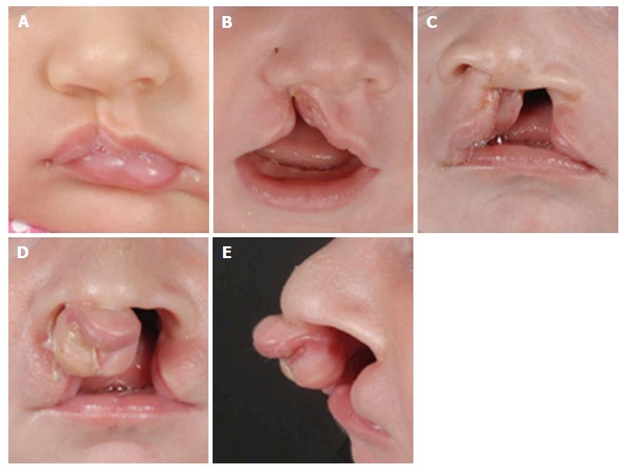
Figure 3 Different types of cleft lips.
A: Unilateral microform cleft lip; B: Unilateral cleft lip and alveolus; C: Unilateral cleft lip and palate; D: Bilateral cleft lip and palate with protrusion of the intermaxillary process; E: Lateral view of bilateral cleft lip and palate.
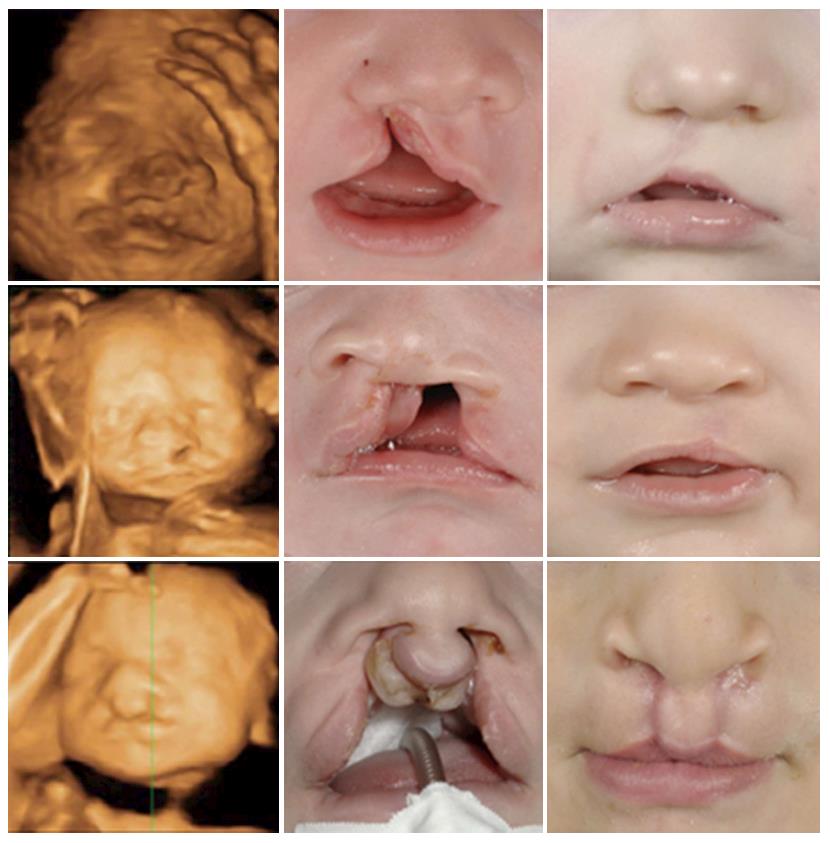
Figure 4 Prenatal, postnatal and post-surgery images of three different patients with cleft lip and alveolus, cleft lip and palate and bilateral cleft lip and palate respectively.
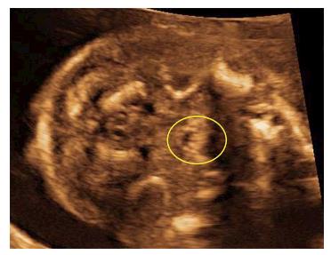
Figure 5 Echo pattern of a normal uvula visualized by ultrasound is typical and strongly resembles an “equals sign”.
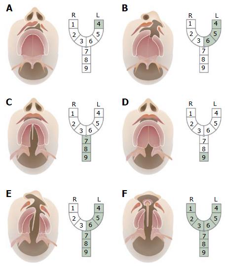
Figure 6 Kernahan’s classification.
The area affected by the cleft is labeled from 1-9, each of which represents a different anatomical structure: 1: Right lip; 2: Right alveolus; 3: Right premaxilla; 4: Left lip; 5: Left alveolus; 6: Left premaxilla; 7: Hard palate; 8: Soft palate; 9: Submucous cleft.
- Citation: Smarius B, Loozen C, Manten W, Bekker M, Pistorius L, Breugem C. Accurate diagnosis of prenatal cleft lip/palate by understanding the embryology. World J Methodol 2017; 7(3): 93-100
- URL: https://www.wjgnet.com/2222-0682/full/v7/i3/93.htm
- DOI: https://dx.doi.org/10.5662/wjm.v7.i3.93


