©2013 Baishideng Publishing Group Co.
World J Methodol. Dec 26, 2013; 3(4): 39-44
Published online Dec 26, 2013. doi: 10.5662/wjm.v3.i4.39
Published online Dec 26, 2013. doi: 10.5662/wjm.v3.i4.39
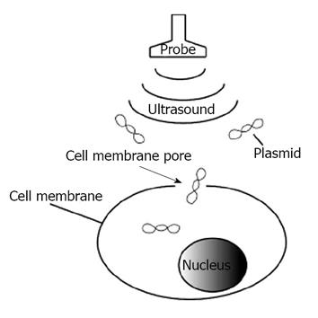
Figure 1 Formation of cell membrane pores after ultrasound irradiation.
Nucleic acid such as plasmids enters the cells through the membrane pores that are formed with ultrasound.
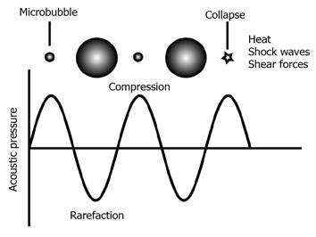
Figure 2 Microbubble response to an ultrasonic pressure wave.
Microbubbles expand and contract when exposed to ultrasound at rarefaction and compression, respectively. At high pressure, microbubbles collapse and a shock wave is emitted.
- Citation: Tomizawa M, Shinozaki F, Motoyoshi Y, Sugiyama T, Yamamoto S, Sueishi M. Sonoporation: Gene transfer using ultrasound. World J Methodol 2013; 3(4): 39-44
- URL: https://www.wjgnet.com/2222-0682/full/v3/i4/39.htm
- DOI: https://dx.doi.org/10.5662/wjm.v3.i4.39


