
















Copyright: ©Author(s) 2026.
World J Methodol. Sep 20, 2026; 16(3): 116523
Published online Sep 20, 2026. doi: 10.5662/wjm.116523
Published online Sep 20, 2026. doi: 10.5662/wjm.116523
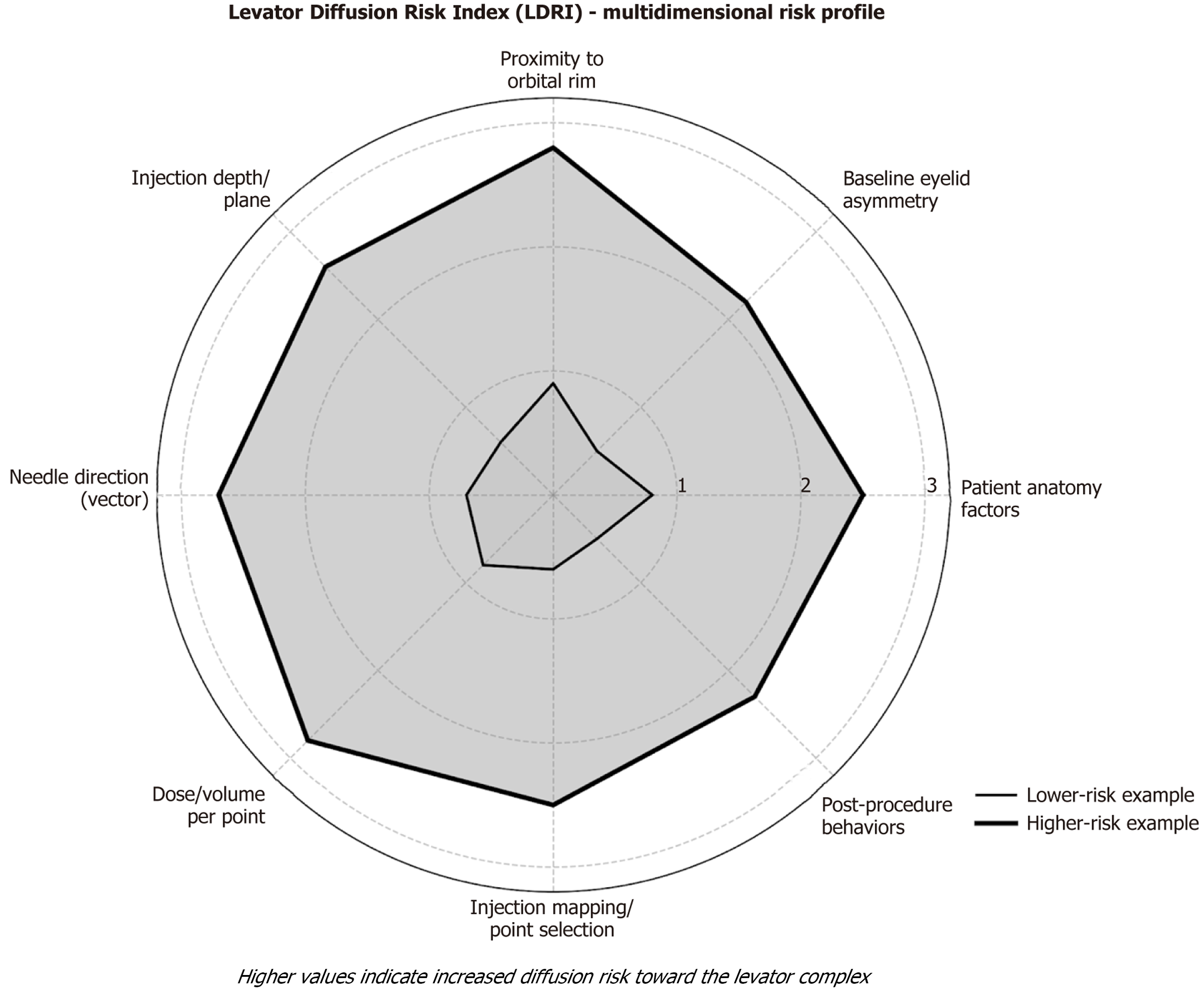
Figure 1 Levator Diffusion Risk Index radar chart.
Radar chart of Levator Diffusion Risk Index domains; overlaid examples show lower vs higher diffusion-risk profiles. Each axis is scored on an ordinal 1-3 scale; higher values indicate increased diffusion risk toward the levator complex. LDRI: Levator Diffusion Risk Index.
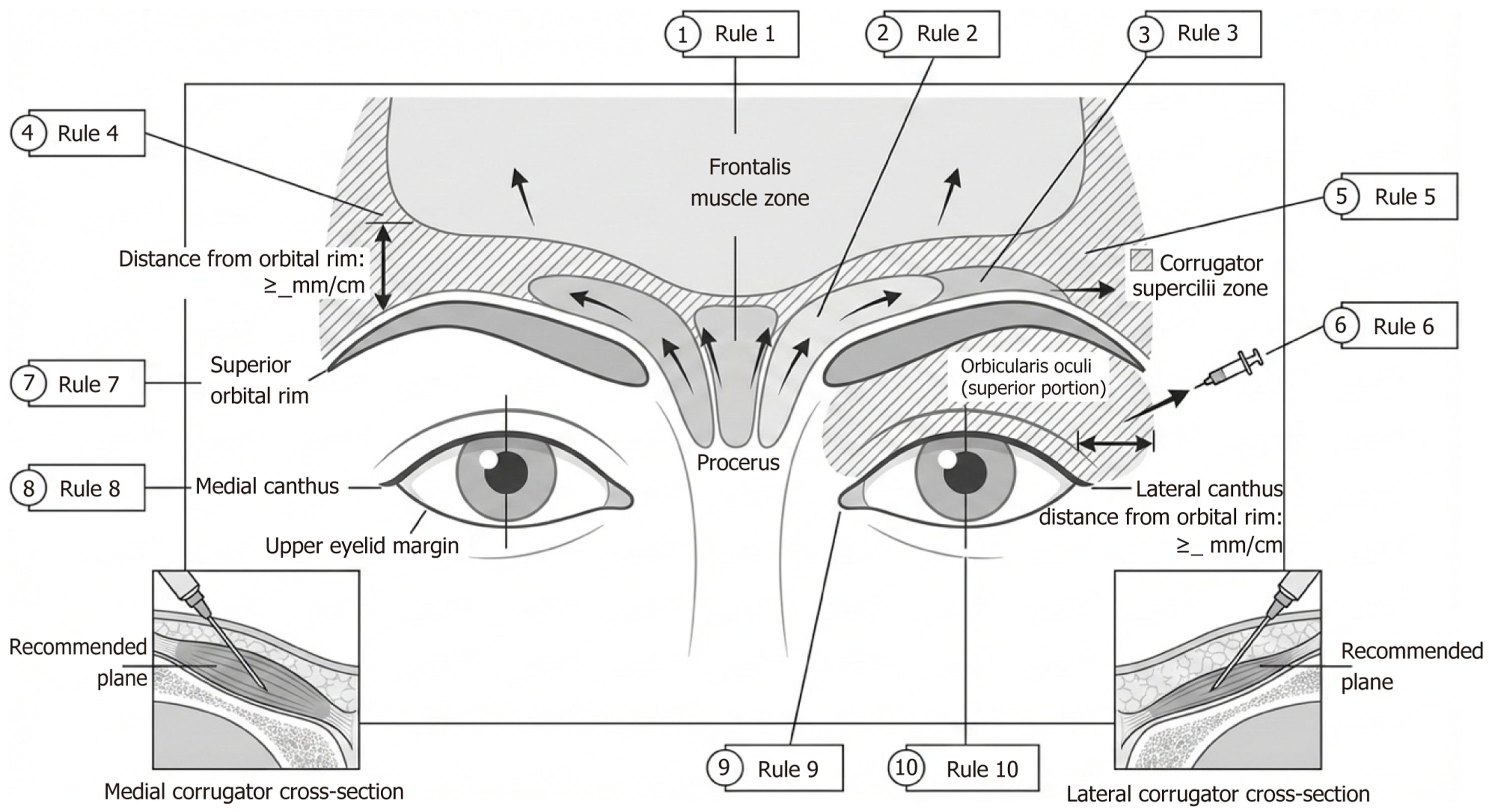
Figure 2 Anatomical schematic of injection safety rules.
Upper-face schematic highlighting the orbital rim boundary, safe zones, needle vectors, and plane guidance; callouts correspond to Box 1.
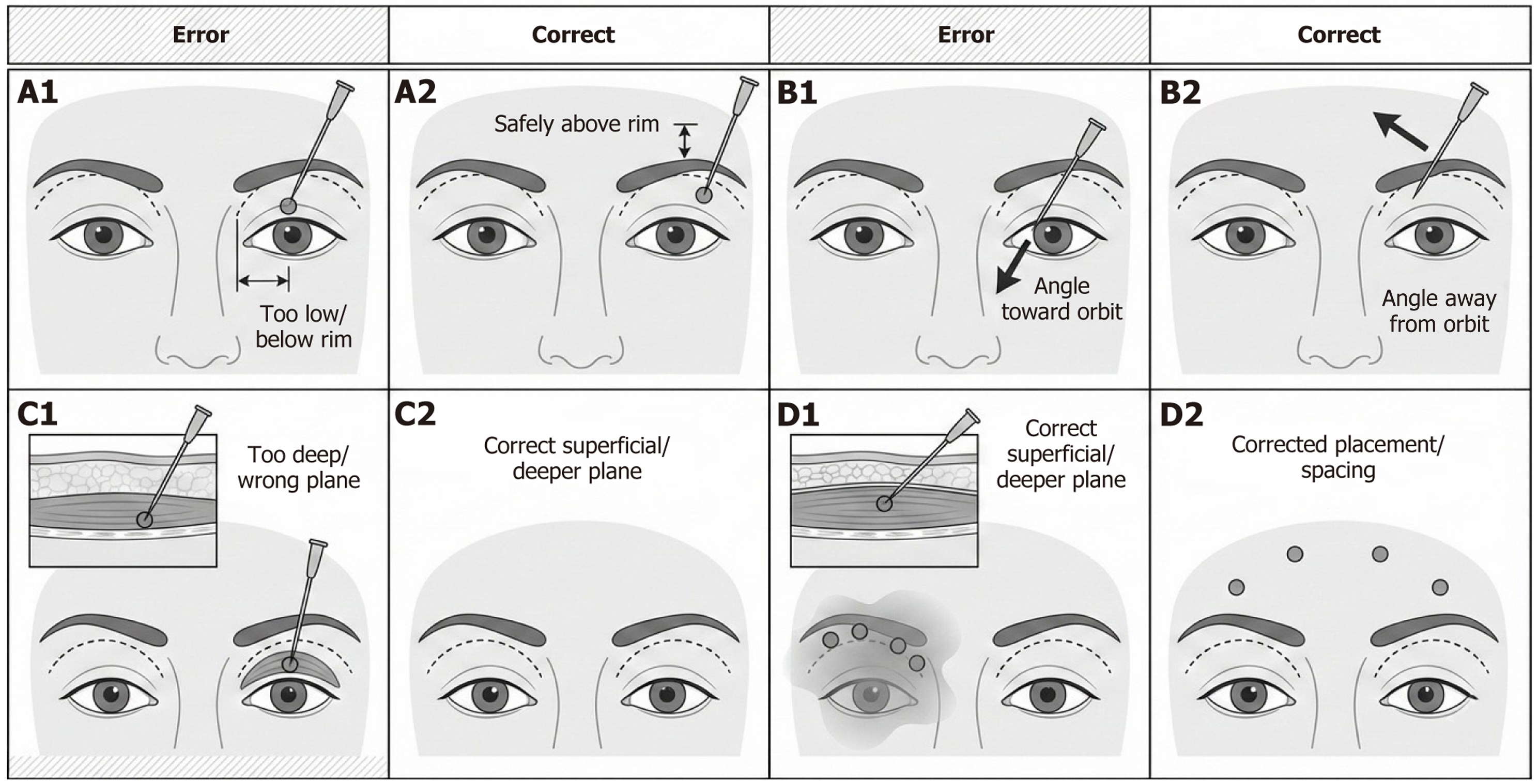
Figure 3 Errors vs corrected injection technique.
Paired schematics contrasting a common technical error (left column, A1, B1, C1, D1) with the corresponding corrected technique (right column, A2, B2, C2, D2) to reduce toxin diffusion toward the levator complex. A1: Injection point placed too close to the orbital rim; A2: Injection point kept superior to the orbital-rim buffer; B1: Needle vector directed toward the orbit; B2: Needle vector directed away from the orbit; C1: Deep placement at the lateral corrugator; C2: Superficial placement at the lateral corrugator; D1: Large per-point volume with clustered points near the rim; D2: Small per-point volumes with adequately spaced points.
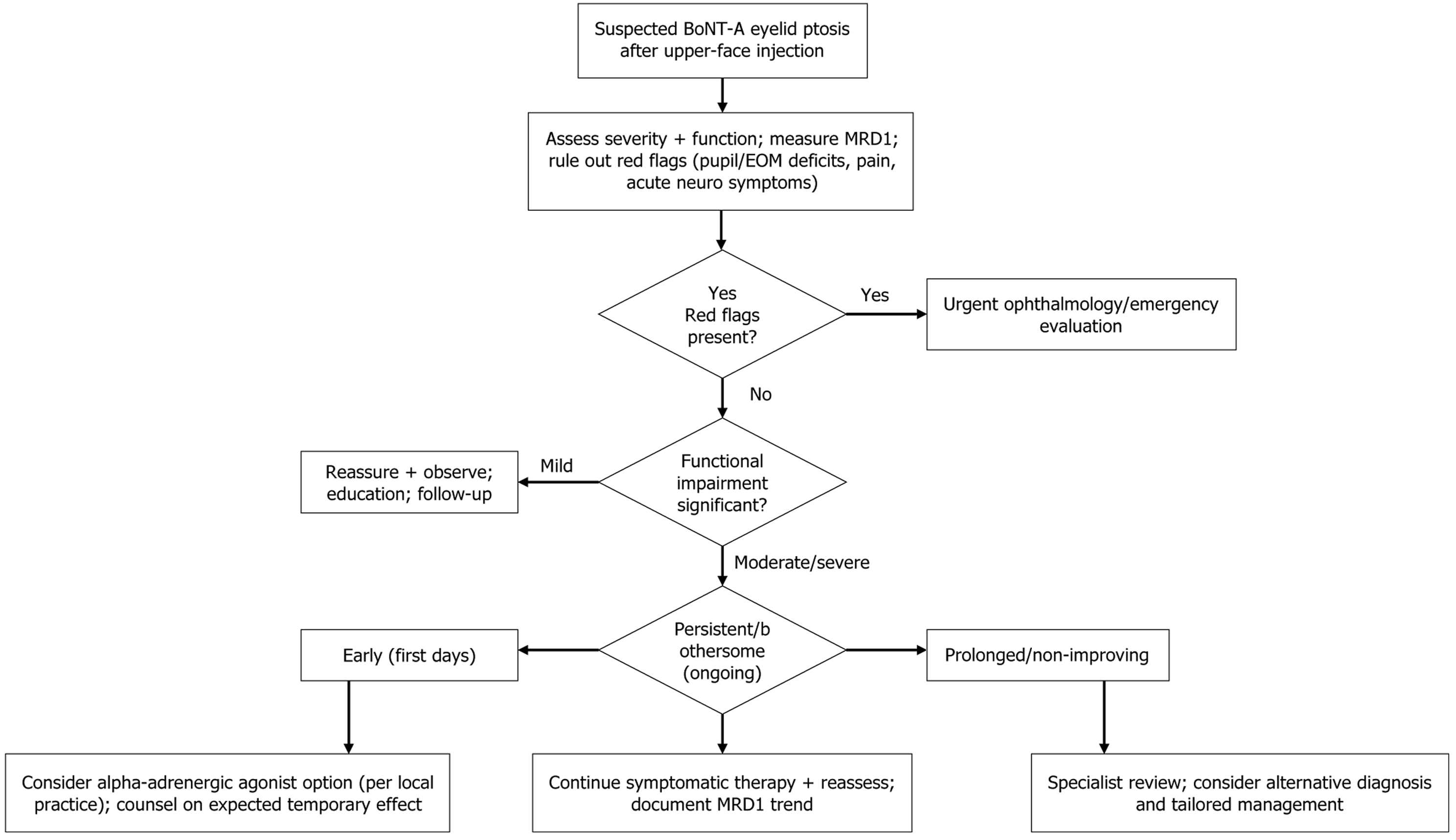
Figure 4 Symptom-onset-anchored management algorithm.
Decision tree for Botulinum toxin A eyelid ptosis based on red-flag screening, functional impact, and time since onset, with stepwise escalation and evidence-grade tags. BoNT-A: Botulinum toxin A; MRD: Margin reflex distance; EOM: Extraocular muscles.
- Citation: Diab N, Bou Farah E, Berjawi A, Ghorayeb T, Beydoun K, Ghanem S, Hamou A, Tarchichi A, Tlaiss Y. Methodology for preventing Botulinum toxin-A eyelid ptosis: Risk index derivation, safety rules, and evidence-graded management algorithm. World J Methodol 2026; 16(3): 116523
- URL: https://www.wjgnet.com/2222-0682/full/v16/i3/116523.htm
- DOI: https://dx.doi.org/10.5662/wjm.116523