Copyright
©The Author(s) 2025.
World J Methodol. Dec 20, 2025; 15(4): 104655
Published online Dec 20, 2025. doi: 10.5662/wjm.v15.i4.104655
Published online Dec 20, 2025. doi: 10.5662/wjm.v15.i4.104655
Figure 1 Shows access cavities.
A: Wrt 11; B: Wrt 12.
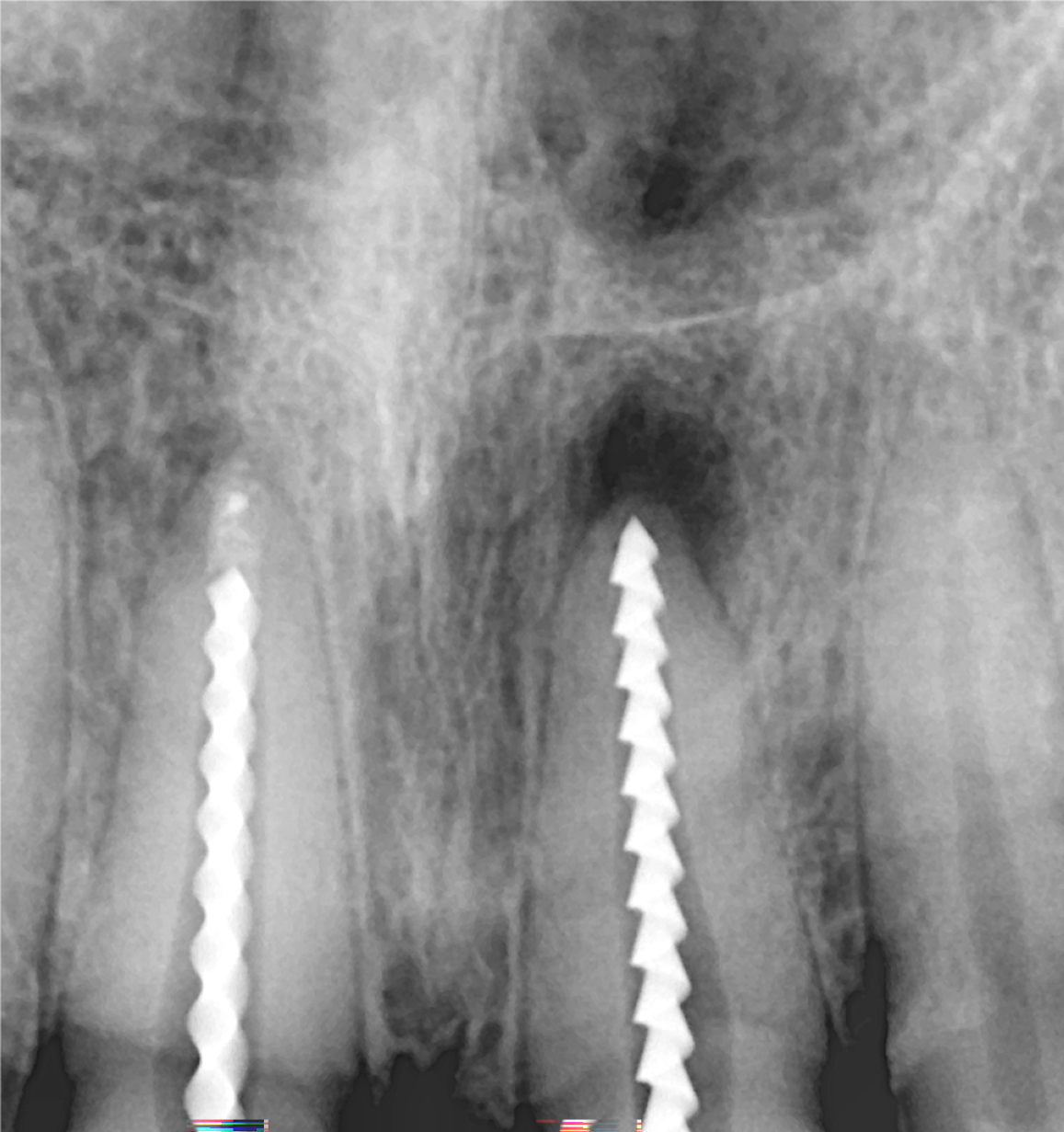
Figure 2 Shows the working length wrt 11 and 21.
Figure 3 Shows the clinal picture of mineral trioxide aggregate placement.
A: Under magnification at 16×; B: Under magnification at 20×.
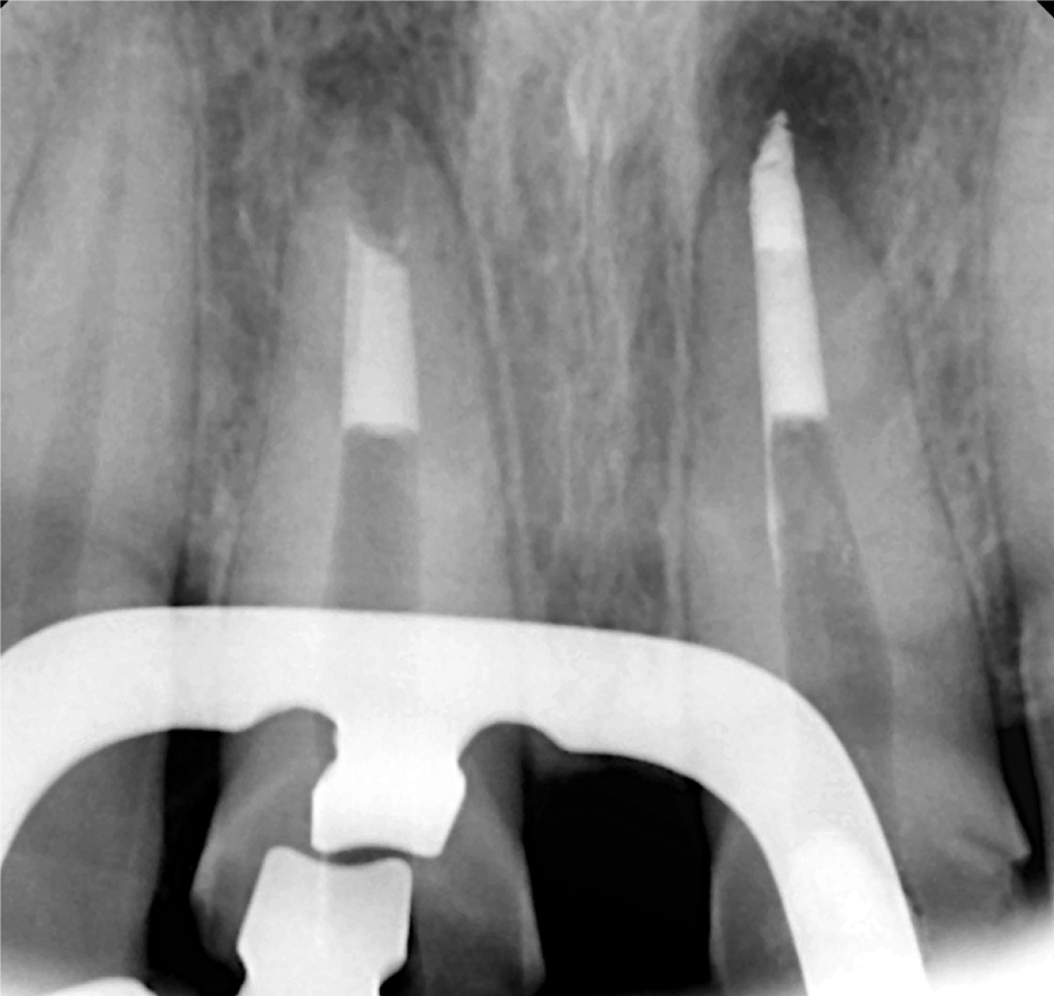
Figure 4 Shows the radiographs of 5 mm mineral trioxide aggregate plug.
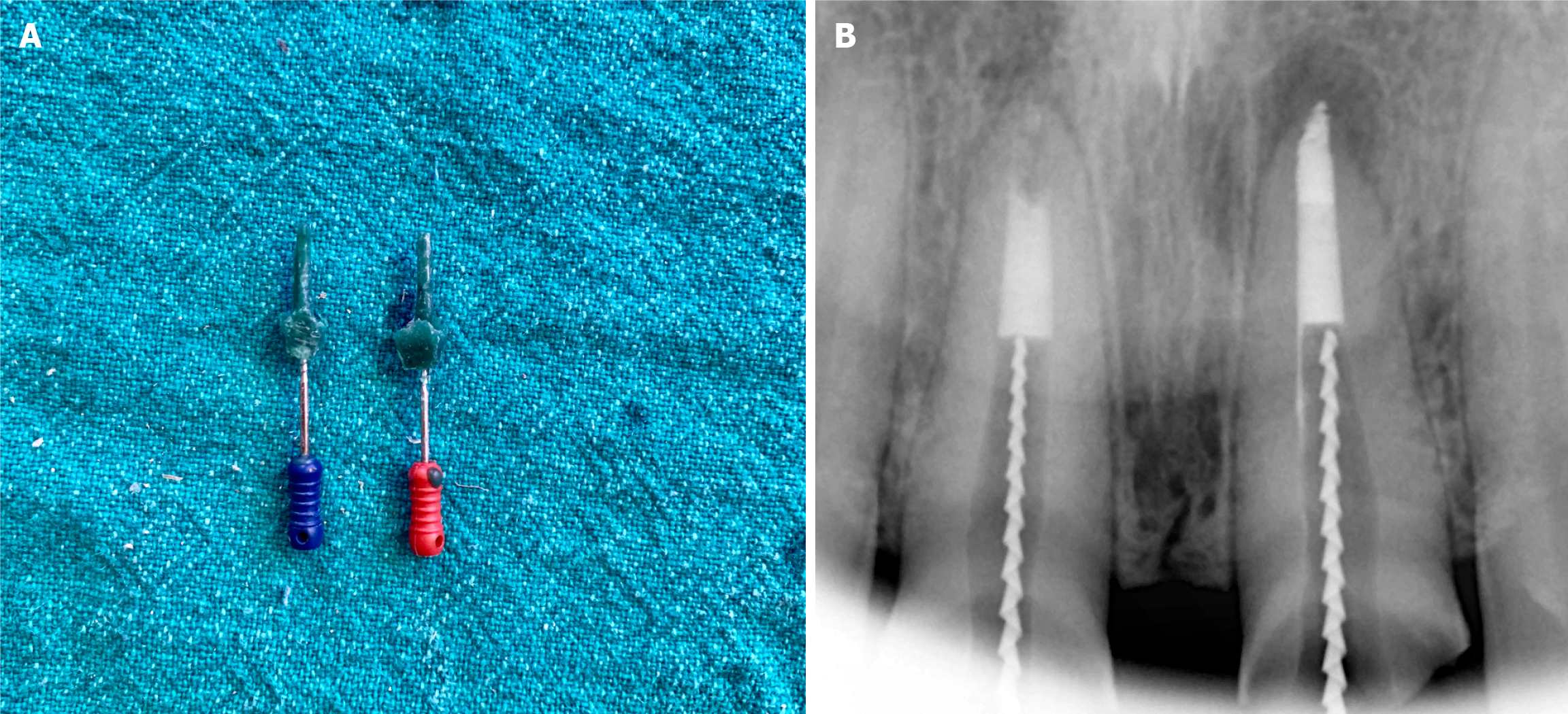
Figure 5 Shows the cast post-impression clinically and radiographically.
A: Clinical pictures show the cast post impressions; B: Radiographically showing show the cast post impressions.
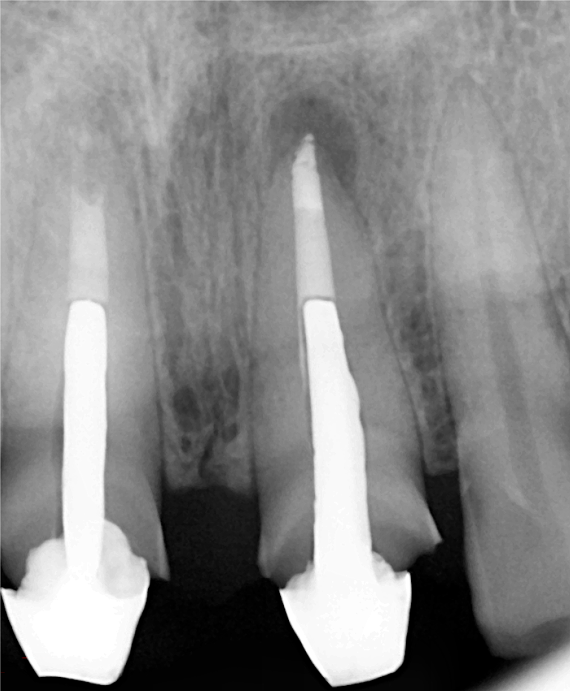
Figure 6 Shows the radiograph of cast post cementation.
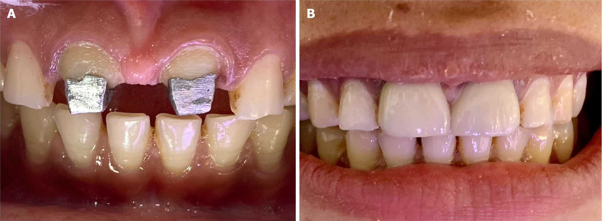
Figure 7 Shows the clinical picture.
A: Cast post cementation; B: zirconia crown cementation
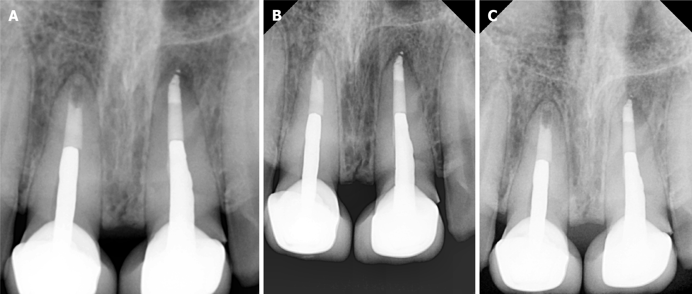
Figure 8 Shows radiograph.
A: 3 months follow up; B: 6 months follow up; C: 1 year follow up.
- Citation: Chauhan R, Chauhan S, Bhasin P, Sood A, Kumar H, Gupta A, Bhasin M. Precision at the apex: Apexification under magnification: A case report. World J Methodol 2025; 15(4): 104655
- URL: https://www.wjgnet.com/2222-0682/full/v15/i4/104655.htm
- DOI: https://dx.doi.org/10.5662/wjm.v15.i4.104655


