Copyright
©The Author(s) 2024.
World J Methodol. Dec 20, 2024; 14(4): 95685
Published online Dec 20, 2024. doi: 10.5662/wjm.v14.i4.95685
Published online Dec 20, 2024. doi: 10.5662/wjm.v14.i4.95685
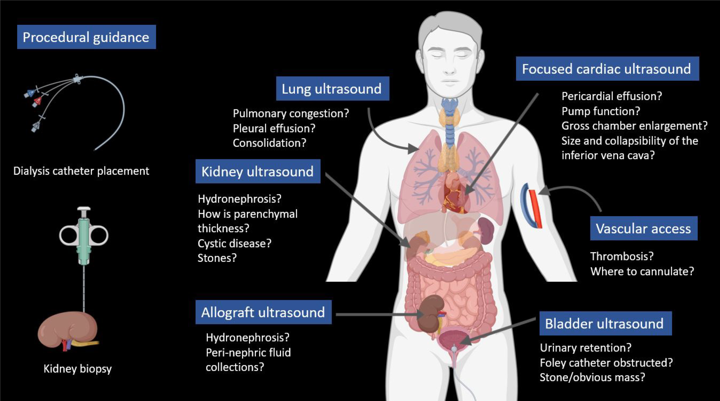
Figure 1 Basic nephrology-related point-of-care ultrasonography: This figure illustrates the organs examined and common clinical questions encountered[2].
Basic POCUS includes greyscale ultrasound and color Doppler but excludes spectral Doppler, which assesses blood flow patterns and velocities. Citation: Koratala A, Kazory A. An Introduction to Point-of-Care Ultrasound: Laennec to Lichtenstein. Adv Chronic Kidney Dis 2021; 28: 193-199. The authors have obtained the permission (Supplementary material).
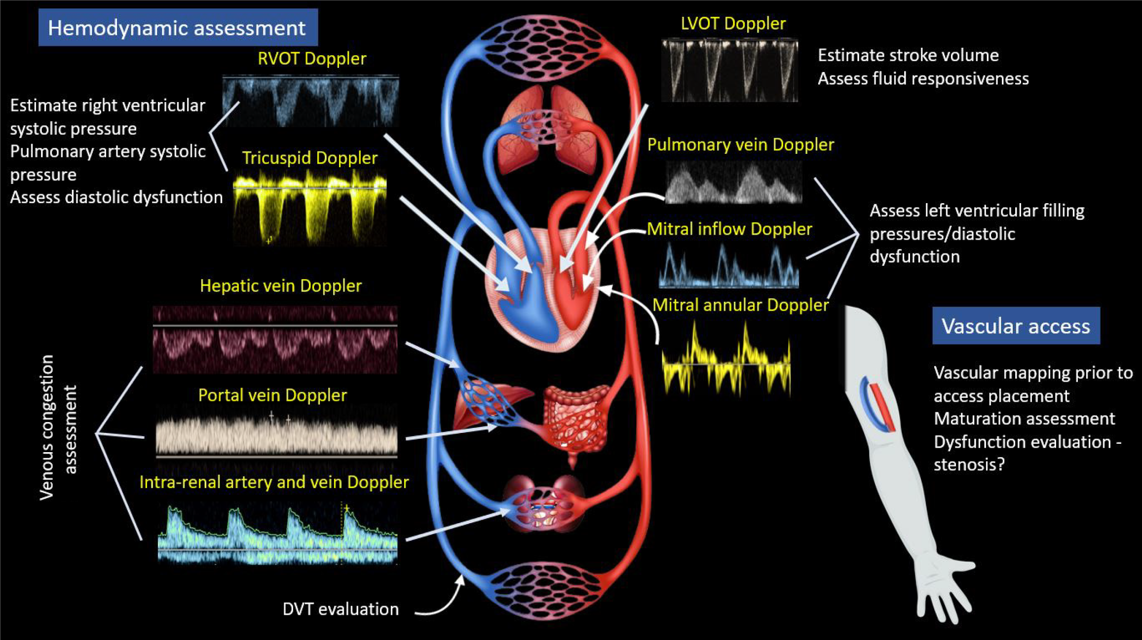
Figure 2 Advanced nephrology-related point-of-care ultrasonography applications involving spectral Doppler[2].
RVOT: Right ventricular outflow tract; LVOT: Left ventricular outflow tract; DVT: Deep vein thrombosis. Citation: Koratala A, Kazory A. An Introduction to Point-of-Care Ultrasound: Laennec to Lichtenstein. Adv Chronic Kidney Dis 2021; 28: 193-199. The authors have obtained the permission (Supplementary material).
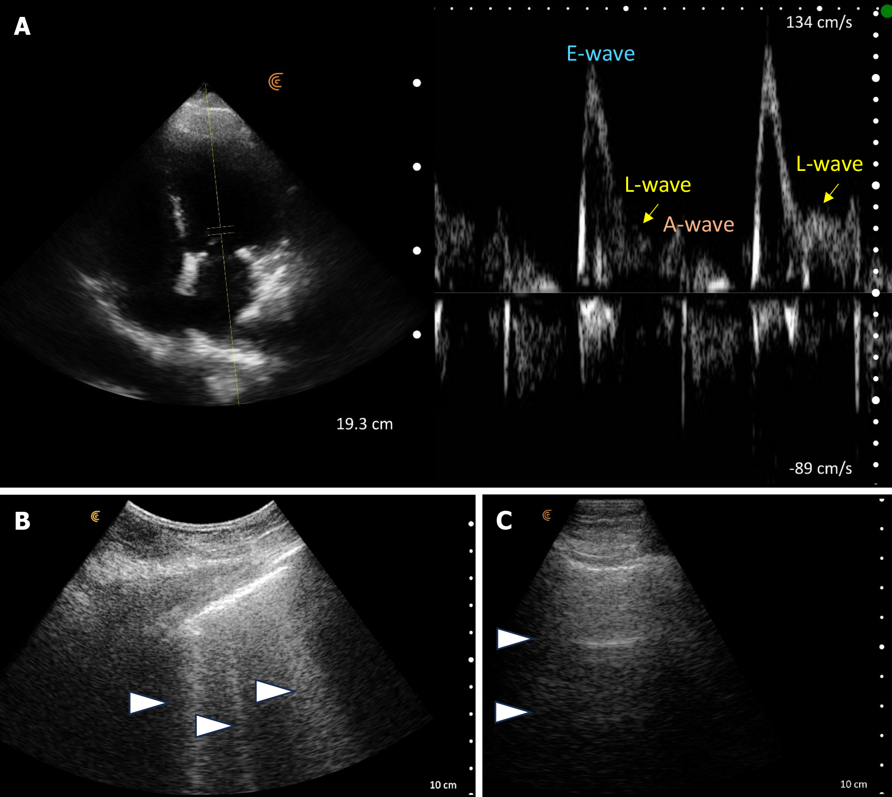
Figure 3 Point-of-care ultrasound images at the time of nephrology consult.
A: An elevated E-wave to A-wave ratio and an L-wave on transmitral Doppler; B: Vertical B-lines on lung ultrasound at initial examination (arrowheads). Follow up lung ultrasound; C: Horizontal A-lines (arrowheads) on lung ultrasound - normal finding.
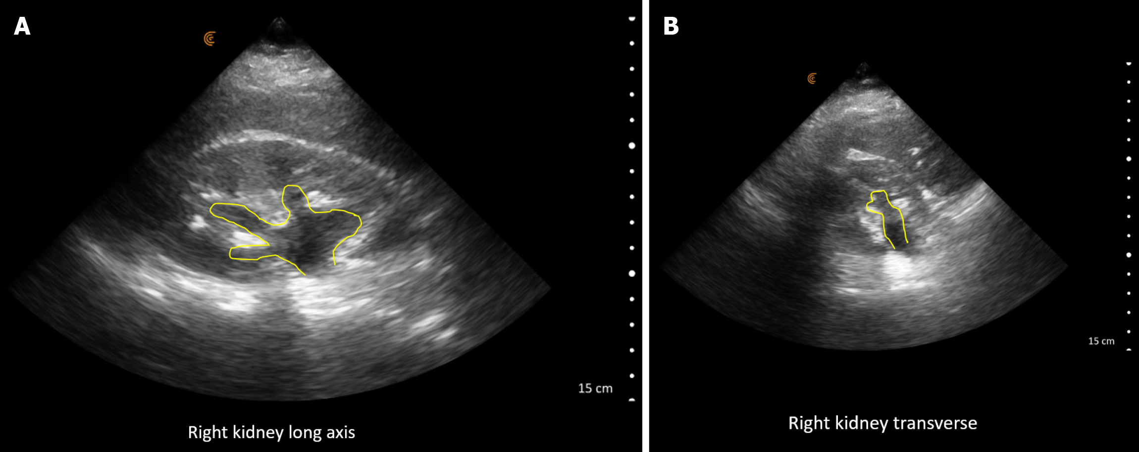
Figure 4 Renal ultrasound images demonstrating.
A: Long axis views of right hydronephrosis (yellow outline); B: Short axis views of right hydronephrosis (yellow outline).
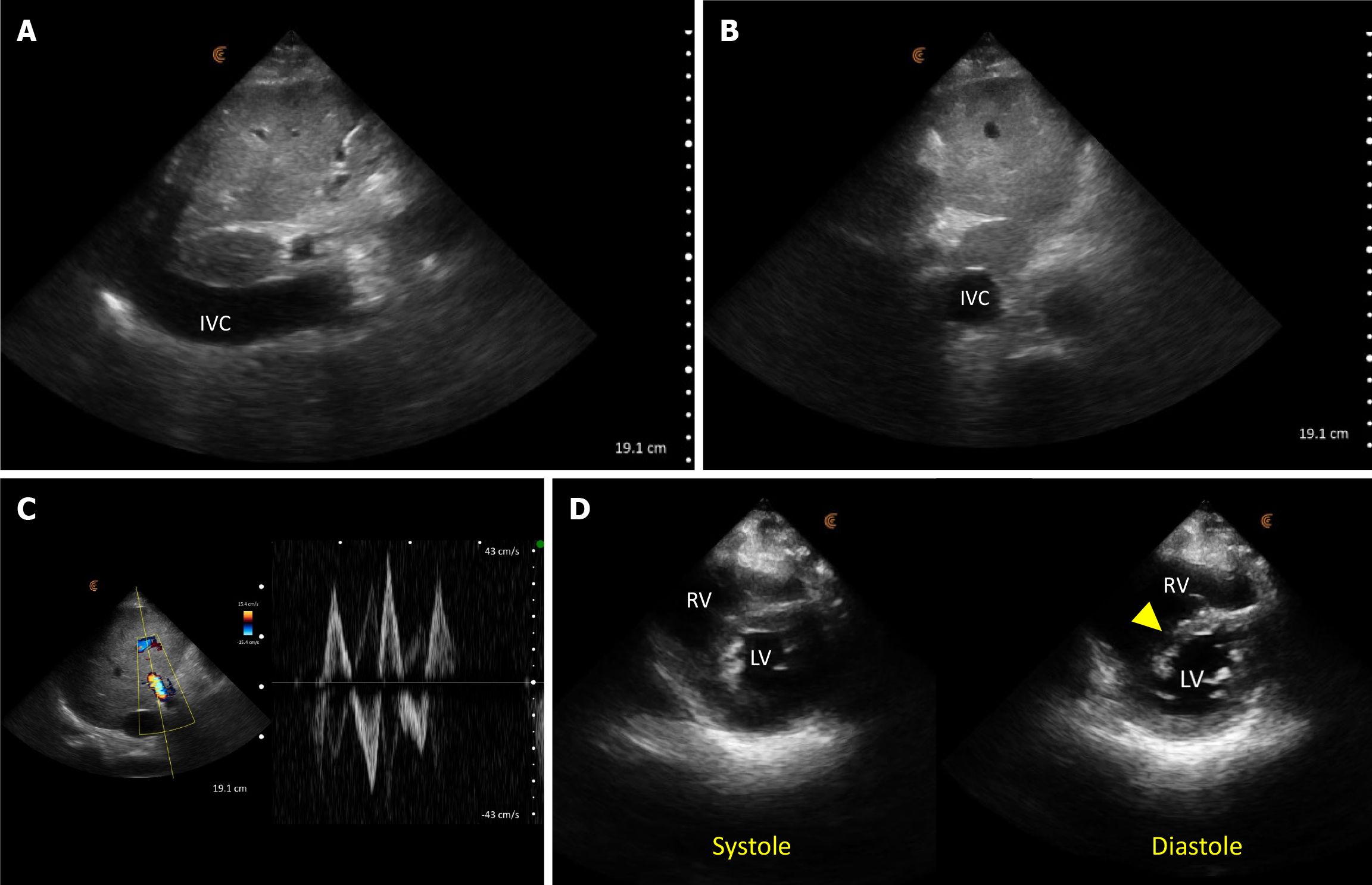
Figure 5 Point-of-care ultrasound images demonstrating findings suggestive of volume/pressure overload.
A: A dilated inferior vena cava (IVC) in long axis; B: IVC transverse view shows a circular vessel as opposed to normal elliptical shape; C: Pulsatile portal vein Doppler with a to-and-fro pattern (normally it’s continuous); D: Parasternal short axis cardiac view showing interventricular septal flattening (arrowhead) predominantly in diastole. RV: Right ventricle; LV: Left ventricle.
Figure 6 Follow up ultrasound examination demonstrating resolution of congestion.
A: The inferior vena cava (indicated by the arrowhead) appearing elliptical in the transverse view with a decreased diameter compared to the initial examination; B: Near-complete collapse of the inferior vena cava (arrowhead) during a sniff, indicative of normal right atrial pressure; C: Continuous waveform observed in the portal vein on Doppler imaging.
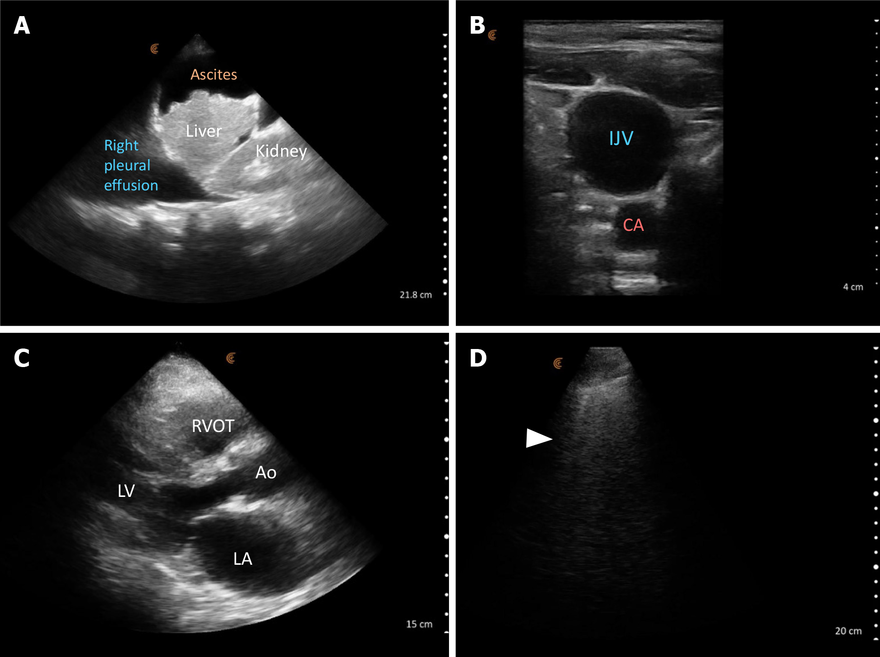
Figure 7 Point-of-care ultrasound images demonstrating findings suggestive of elevated cardiac filling pressures.
A: Right pleural effusion and ascites as seen on right upper quadrant lateral scan plane; B: A dilated internal jugular vein (IJV) at the cricoid cartilage level with a head angle of approximately 45 degrees consistent with elevated right atrial pressure. Adjacent carotid artery (CA) is seen: C: Parasternal long axis cardiac view showing a qualitatively dilated left atrium; D: Lung ultrasound image demonstrating vertical B-lines (arrowhead) indicative of elevated extravascular lung water. LA: left atrium; Ao: Aorta; LV: Left ventricle; RVOT: Right ventricular outflow tract.
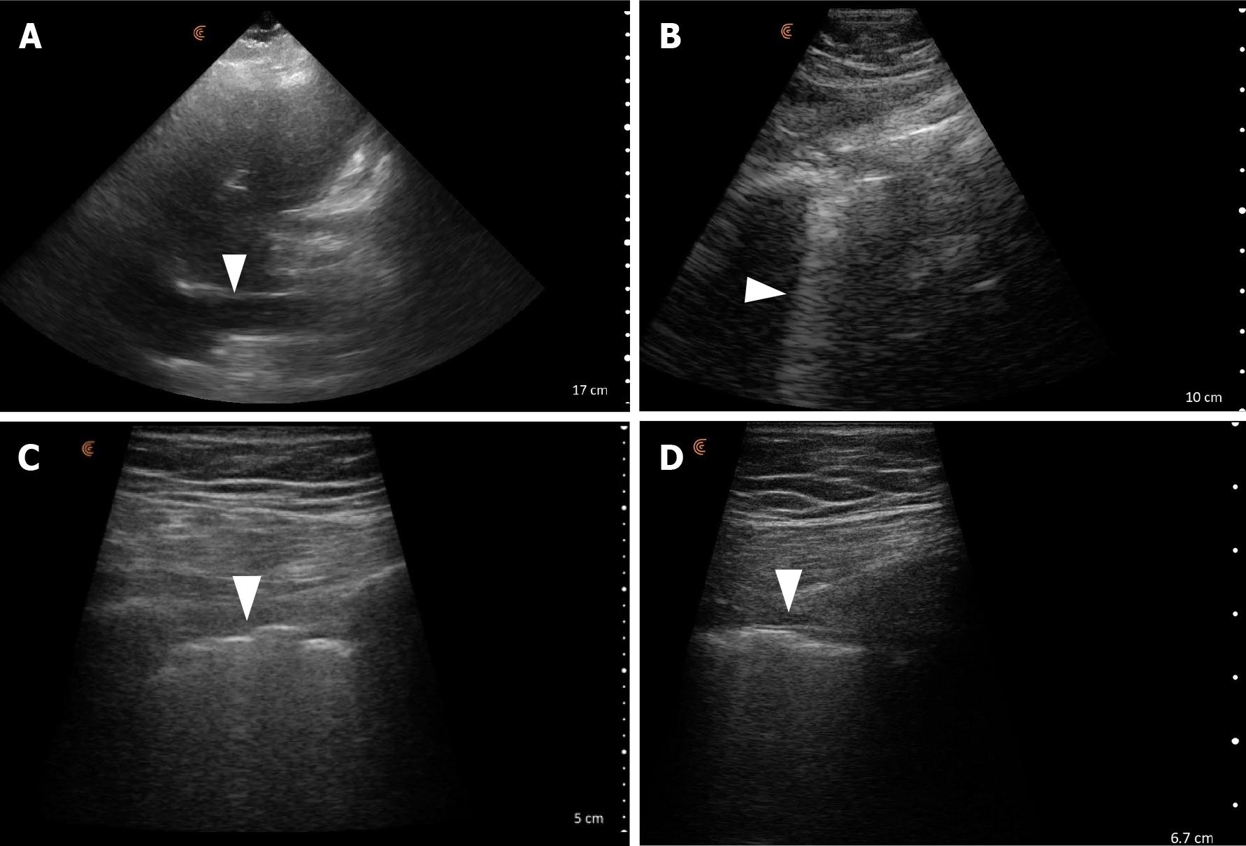
Figure 8 Point-of-care ultrasound findings favoring primary lung pathology over congestive heart failure.
A: A relatively small IVC (< 1.5 cm maximal diameter); B: Confluent B-lines on lung ultrasound (arrowhead); C and D: Irregular pleural line imaged using a high frequency-low depth setting.
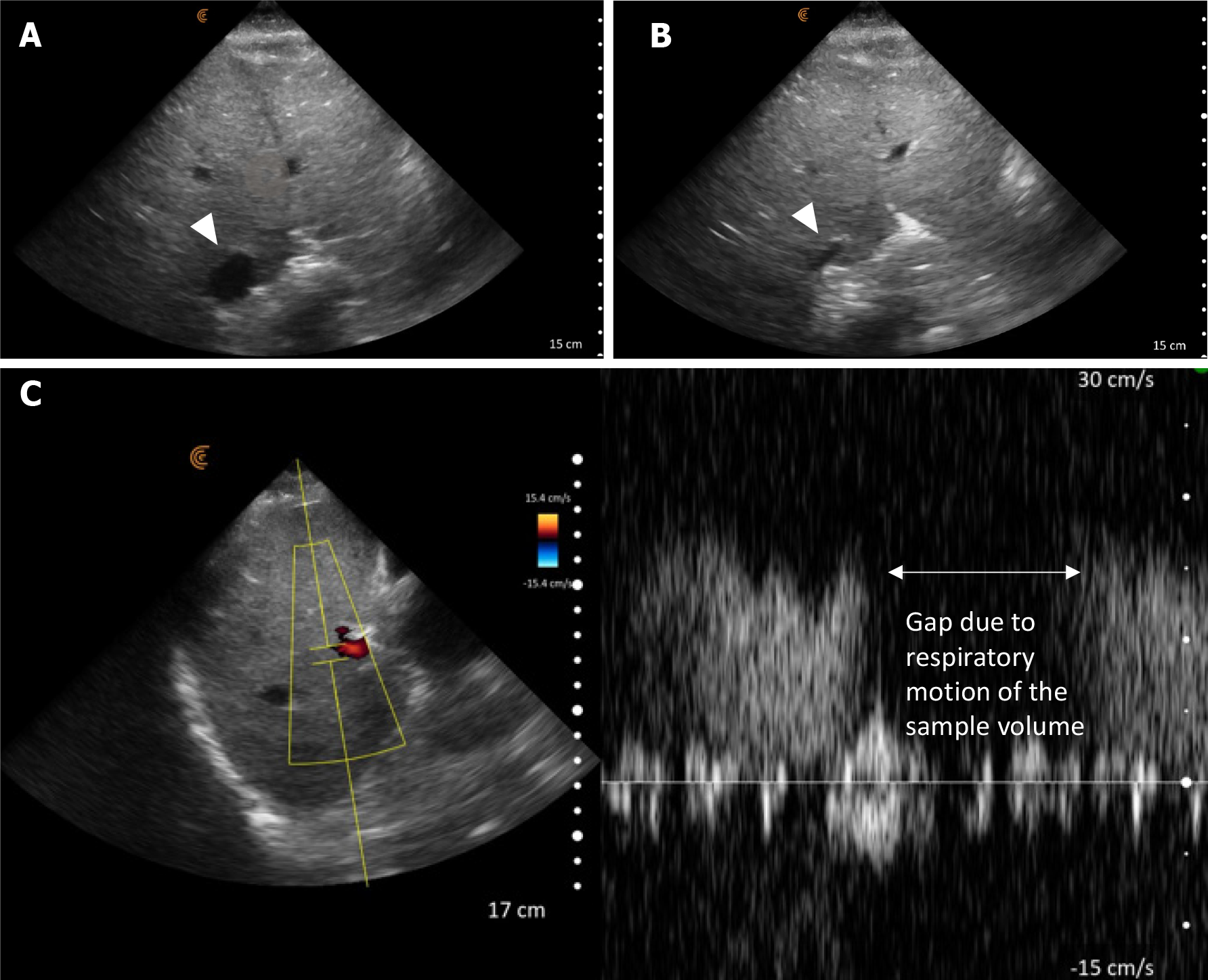
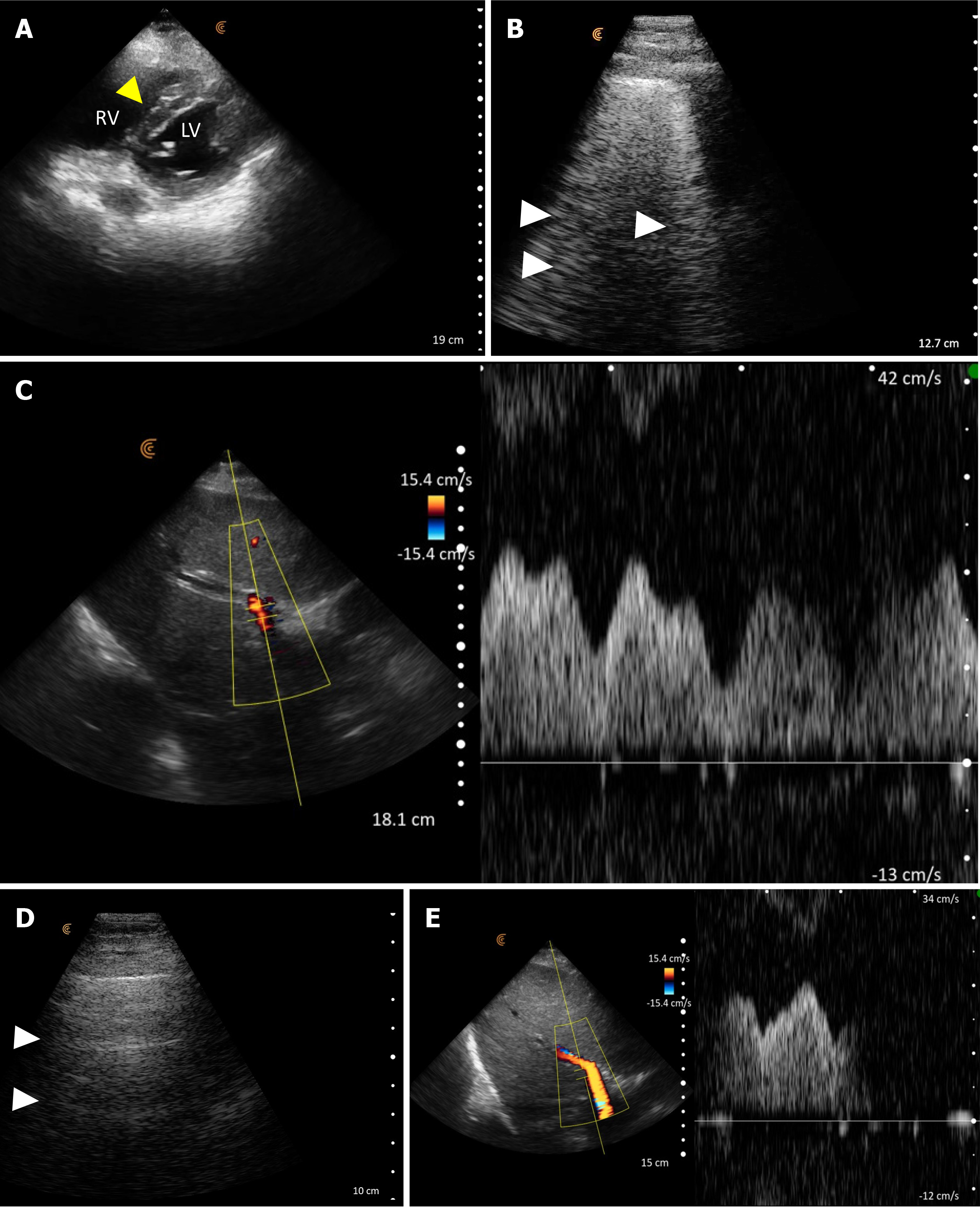
Figure 9 Point-of-care ultrasound images demonstrating stigmata of elevated cardiac filling pressures at the time of nephrology consult.
A: Interventricular septal flattening (arrowhead) on parasternal short axis cardiac view suggestive of elevated right heart pressures; B: B-lines on lung ultrasound (arrowheads); C: Increased portal vein pulsatility > 50% (asterisks represent highest and lowest points in one cardiac cycle); D and E: Represent follow up examination. Normal horizontal A-lines seen on lung ultrasound (D), improved portal vein pulsatility (E).
- Citation: Sinanan R, Moshtaghi A, Koratala A. Point-of-care ultrasound in nephrology: A private practice viewpoint. World J Methodol 2024; 14(4): 95685
- URL: https://www.wjgnet.com/2222-0682/full/v14/i4/95685.htm
- DOI: https://dx.doi.org/10.5662/wjm.v14.i4.95685


