Copyright
©The Author(s) 2022.
World J Methodol. Mar 20, 2022; 12(2): 64-82
Published online Mar 20, 2022. doi: 10.5662/wjm.v12.i2.64
Published online Mar 20, 2022. doi: 10.5662/wjm.v12.i2.64
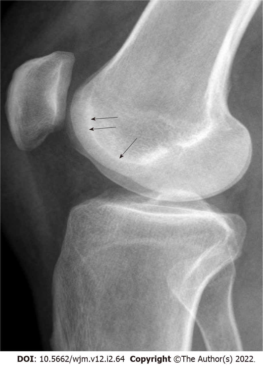
Figure 1 Lines and contours seen in normal people on true lateral radiography.
Anteriorly, parallel dense lines belonging to both condyles and linear density of the base of trochlear sulcus (arrows) just posteriorly are observed. These lines do not intersect with each other. There is no bump or prominence on the anterior aspect.
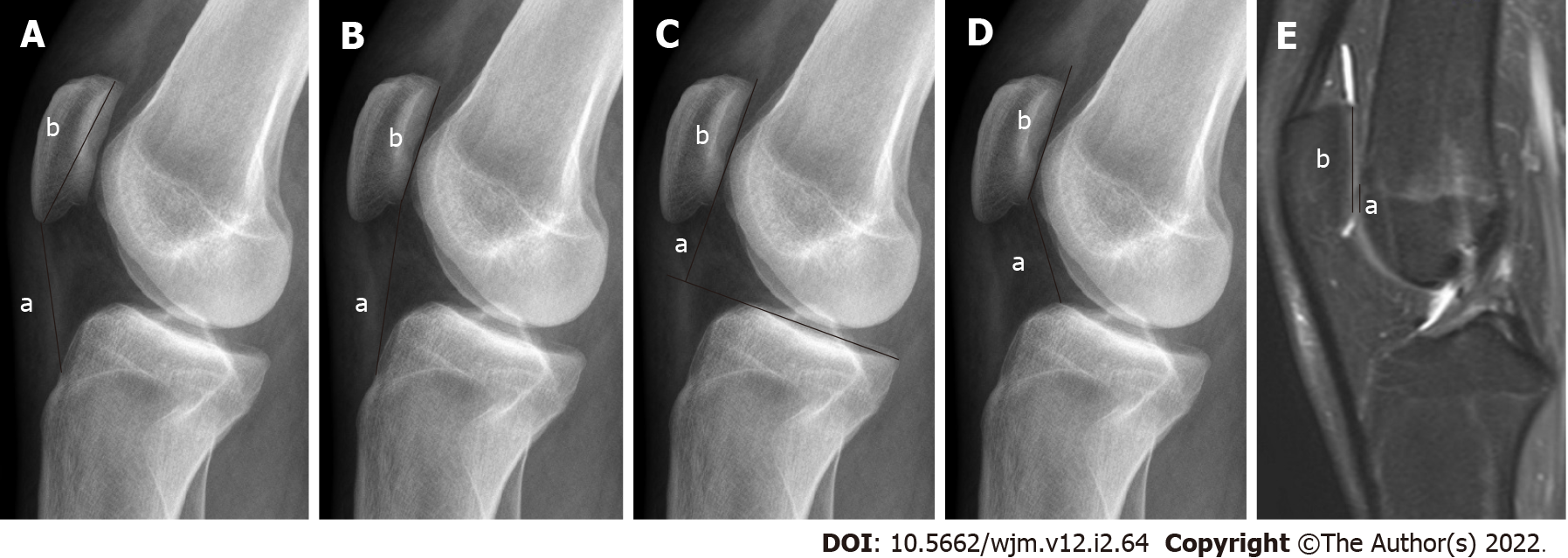
Figure 2 Radiography.
A: Insall-Salvati (IS) ratio (a: Patellar tendon length; b: Length of the patella); B: Modified IS ratio (a: The distance between the lower end of the articular face of the patella and TT; b: Length of the articular face of the patella); C: Blackburne-Peel ratio (a: The distance between the tibial plateau line and the lower pole of the patella joint surface; b: Length of the articular surface of the patella); D: Caton-Deschamps ratio (a: The distance between the inferior point of the patellar articular face and the anterior superior border of the tibia; b: Length of the articular surface of the patella); E: Magnetic resonance imaging, Patellotrochlear index (a: Length of the trochlear cartilage; b: Length of the patellar cartilage). These images are showing different measurement methods for evaluating the patellar height.
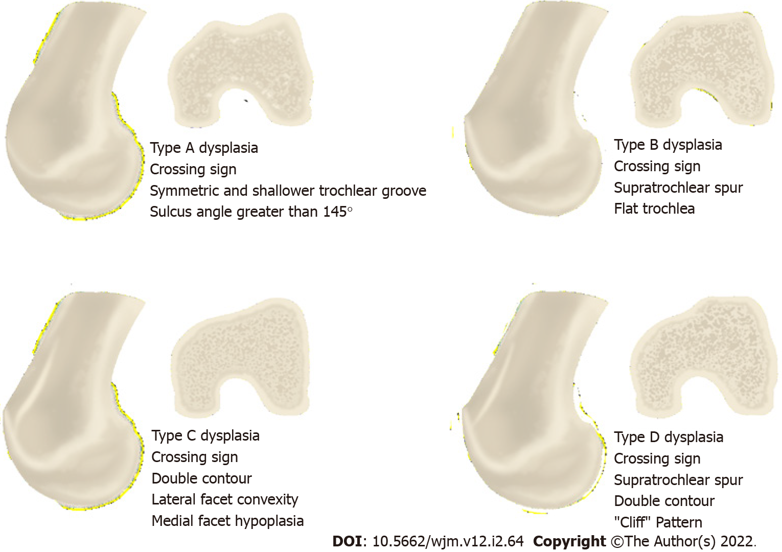
Figure 3 Illustration showing the Dejour classification used in trochlear dysplasia in true lateral radiography and axial slice images.
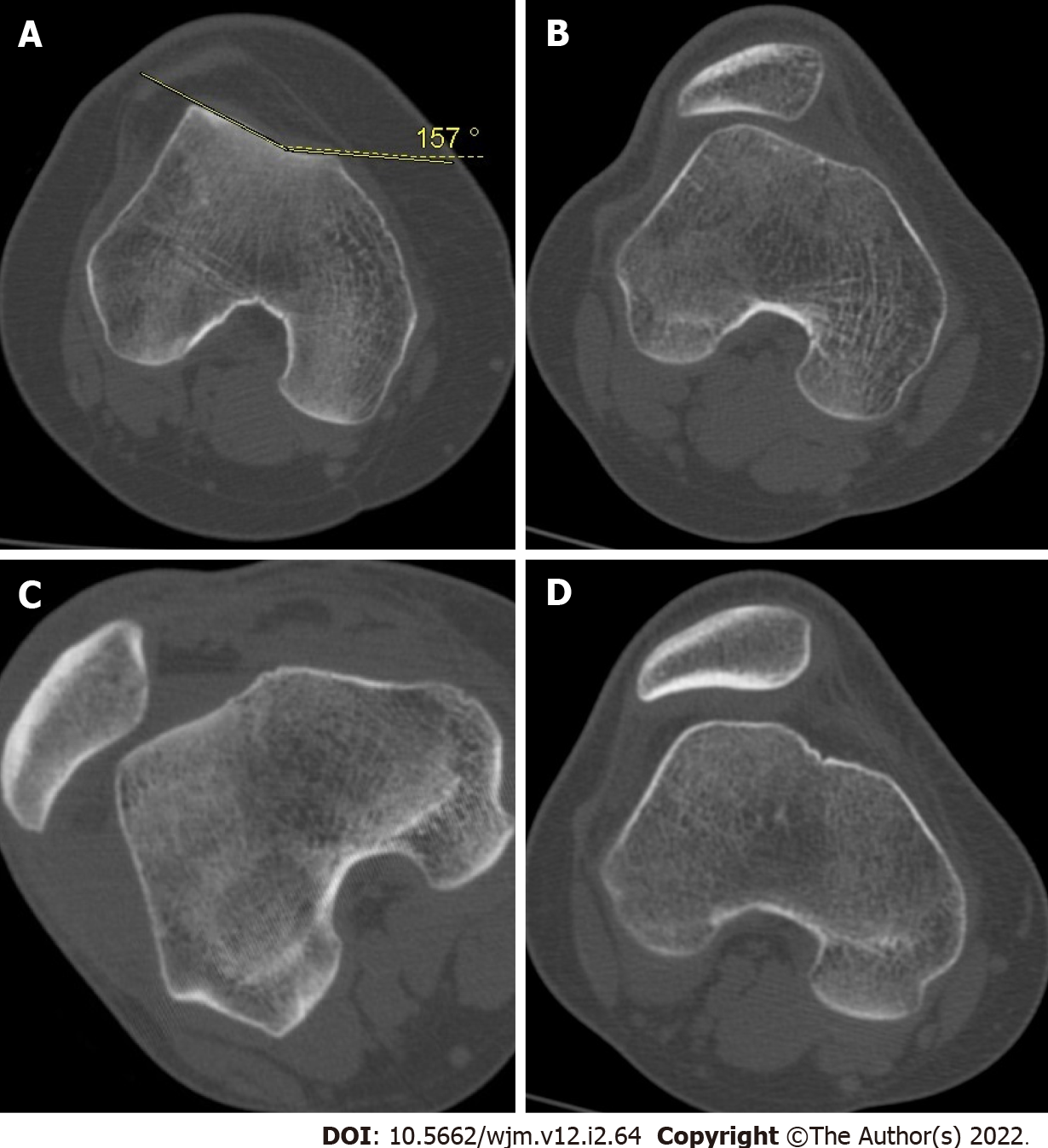
Figure 4 Sample cases with trochlear dysplasia according to Dejour classification.
A: Type A; B: Type B; C: Type C; D: Type D.
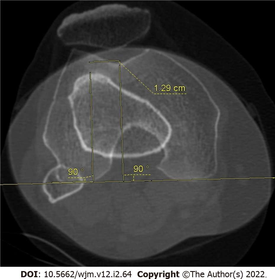
Figure 5 Tibial tuberosity-trochlear groove distance measurement.
Superimposed image of the trochlear groove and tibial tuberosity used in the axial images on computed tomography, and here the lateral offset of tibial tuberosity is evaluated.
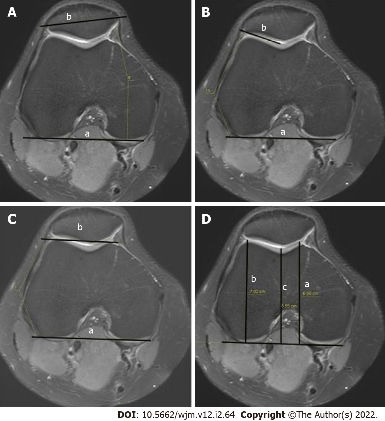
Figure 6 Measurement.
A: Patellar tilt (a: The line joining the posterior femoral condyles; b: The transverse patellar axis); B: Lateral trochlear inclination (a: The line tangent to the posterior edge of the femoral condyles; b: The line drawn tangent to the lateral facet); C: Trochlear angle (a: the angle between the line passing posteriorly to both femoral condyles; b: The line joining the foremost parts of the medial and lateral facets); D: Trochlear depth in axial magnetic resonance imaging (a: The maximal anterior-posterior diameter of the medial femoral condyle; b: The maximal anterior-posterior diameter of the lateral condyle; c: The minimum distance of the deepest point of the trochlea).
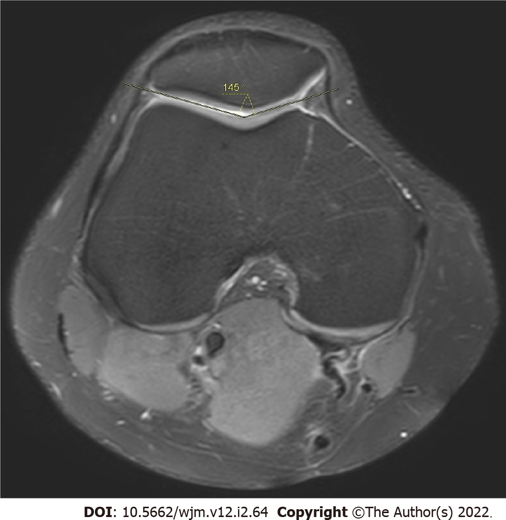
Figure 7 Measurement of sulcus angle.
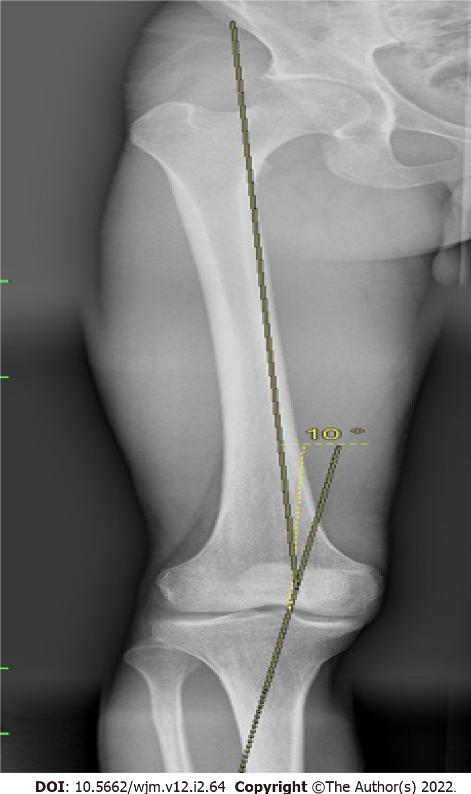
Figure 8 Q angle measurement.
- Citation: Ormeci T, Turkten I, Sakul BU. Radiological evaluation of patellofemoral instability and possible causes of assessment errors. World J Methodol 2022; 12(2): 64-82
- URL: https://www.wjgnet.com/2222-0682/full/v12/i2/64.htm
- DOI: https://dx.doi.org/10.5662/wjm.v12.i2.64


