Copyright
©The Author(s) 2021.
World J Methodol. Jul 20, 2021; 11(4): 110-115
Published online Jul 20, 2021. doi: 10.5662/wjm.v11.i4.110
Published online Jul 20, 2021. doi: 10.5662/wjm.v11.i4.110
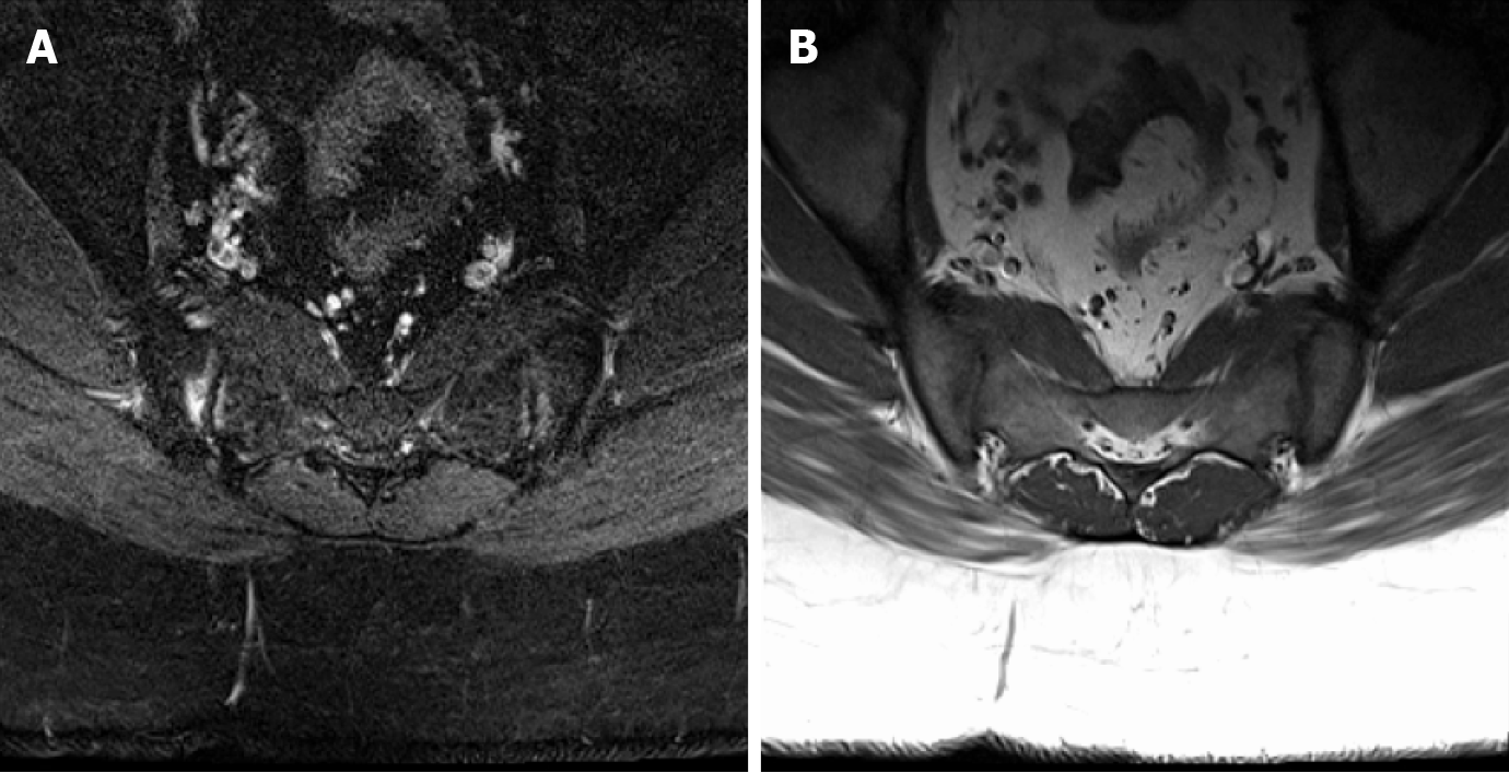
Figure 1 Axial T1 and STIR images demonstrate bilateral sacroiliac joint edema and irregularity consistent with sacroiliitis, considered a major change in diagnosis.
These inflammatory changes resulted in no changes to management in this patient who eventually underwent microdiscectomy for disc extrusion seen on concurrent lumbar spine magnetic resonance. A: Axial T1 image; B: STIR image.
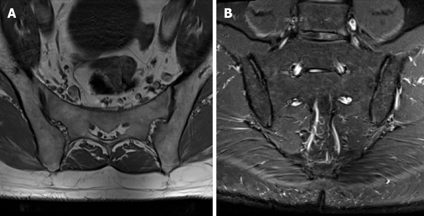
Figure 2 Axial T1 and coronal STIR images demonstrate mild bilateral sacroiliac joint degeneration.
This patient was also noted to have incidentally noted lower lumbar spine degeneration, for which he subsequently underwent a dedicated lumbar spine magnetic resonance. A: Axial T1 image; B: Coronal STIR image.
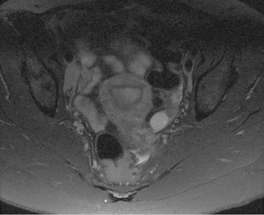
Figure 3 Axial STIR image demonstrates an incidentally noted small left ovarian cyst and borderline enlarged right external iliac lymph nodes in this reproductive age patient with an underlying systemic illness.
No musculoskeletal abnormalities were present on her exam.
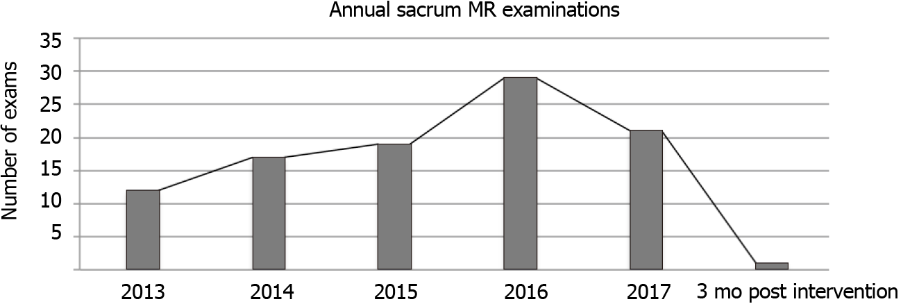
Figure 4 Number of sacral magnetic resonance examinations performed per year during the retrospective review followed by 3 mo post intervention.
MR: Magnetic resonance.
- Citation: Castillo S, Joodi R, Williams LE, Pezeshk P, Chhabra A. Sacrum magnetic resonance imaging for low back and tail bone pain: A quality initiative to evaluate and improve imaging utility. World J Methodol 2021; 11(4): 110-115
- URL: https://www.wjgnet.com/2222-0682/full/v11/i4/110.htm
- DOI: https://dx.doi.org/10.5662/wjm.v11.i4.110


