
















Copyright: ©Author(s) 2026.
World J Transl Med. Jul 28, 2026; 12(2): 122418
Published online Jul 28, 2026. doi: 10.5528/wjtm.122418
Published online Jul 28, 2026. doi: 10.5528/wjtm.122418
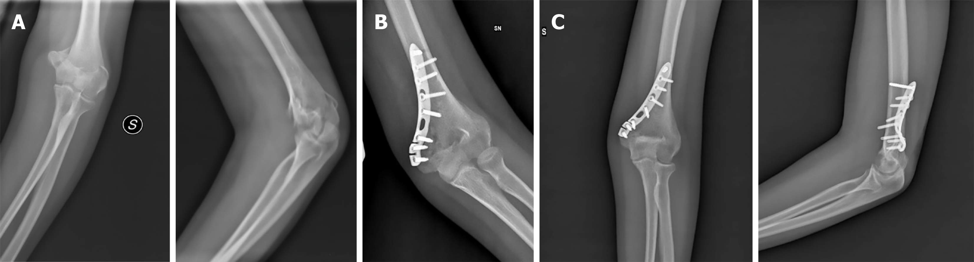
Figure 1 Case 1.
A: Anteroposterior and lateral radiographs, taken at the time of admission, showing an articular distal humeral fracture of a 29-year-old man; B: Postoperative radiograph taken after open reduction and internal fixation with a medial plate. The patient developed symptoms of acute fracture-related infection (local redness, pain and a fistula) 20 days after the open reduction and internal fixation; C: Healed fracture 8 weeks after a debridement, antibiotics and implant retention (debridement, antibiotics, and implant retention procedure).
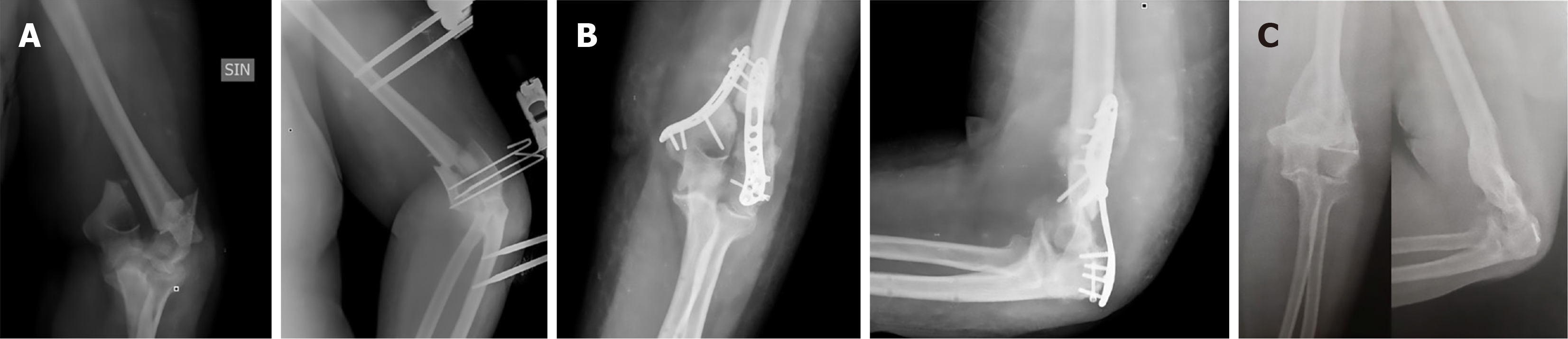
Figure 2 Case 2.
A: Anteroposterior radiographs, taken at the time of admission, showing a displaced open articular distal humerus fracture and temporary stabilization with K-wires and an external fixator. After 10 days, open reduction and internal fixation was performed using two perpendicular distal humeral plates; B: Non-union with hardware failure 3 months after surgery, associated to high erythrocyte sedimentation rate and C-reactive protein blood levels; C: Fracture union after an implant removal and prolonged immobilization in a static posterior splint.
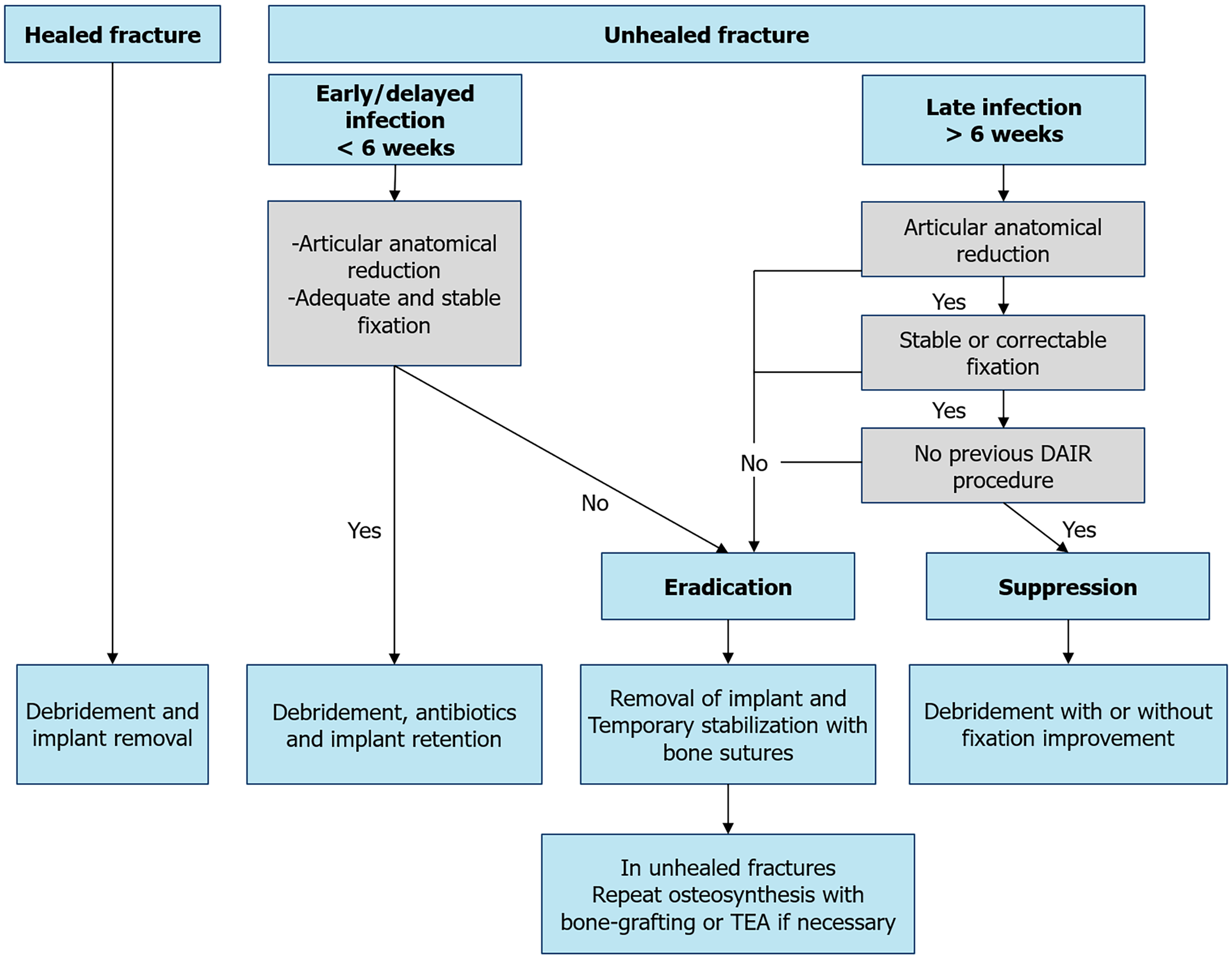
Figure 3 Our treatment strategy for distal humerus fracture-related infections.
TEA: Total elbow arthroplasty.
- Citation: Bardellini G, Rodà D, Abate B, Celli A. Management of distal humerus fracture-related infections after fixation: A retrospective study. World J Transl Med 2026; 12(2): 122418
- URL: https://www.wjgnet.com/2220-6132/full/v12/i2/122418.htm
- DOI: https://dx.doi.org/10.5528/wjtm.122418