
















Copyright: ©Author(s) 2026.
World J Transl Med. Jul 28, 2026; 12(2): 122417
Published online Jul 28, 2026. doi: 10.5528/wjtm.122417
Published online Jul 28, 2026. doi: 10.5528/wjtm.122417
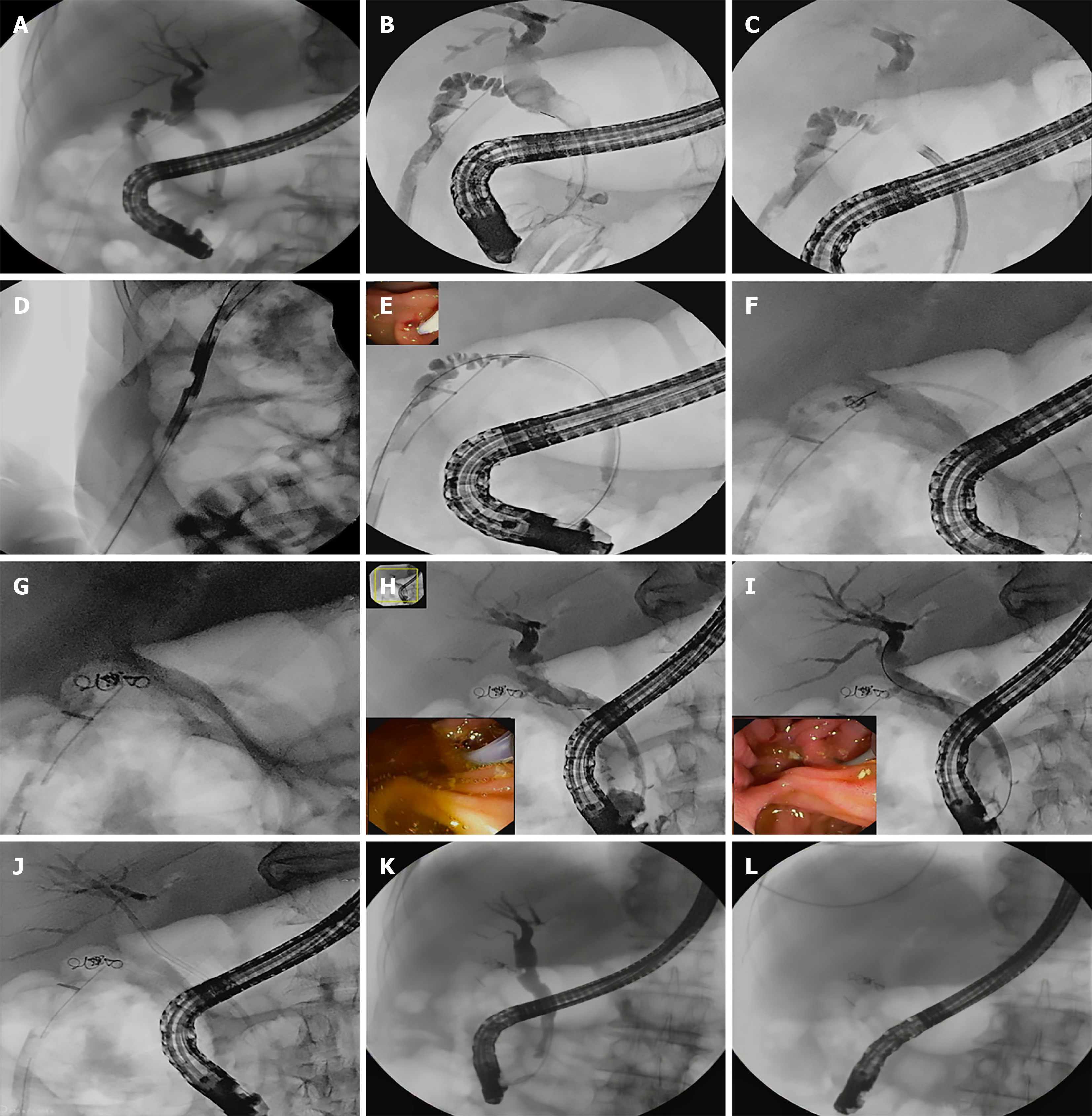
Figure 1 Imaging results.
A: Cystic duct leak with percutaneous drain in gallbladder fossa; B: Biliary cannulation with sphincterotome tip at cystic duct opening; C: SpyGlass cholangioscopy guided cannulation of the cystic duct stump; D: Guidewire placement into the cystic duct via the existing percutaneous drainage catheter; E: Cannula was advanced over the guidewire through the percutaneous drainage tube for selective cystic duct cannulation (panel inset endoscopy showing cannulation); F: Initial deployment of the first embolization coil within the cystic duct stump, viewed under fluoroscopic guidance; G: Fluoroscopic image confirming placement of three embolization coils in the cystic duct stump; H and I: Cholangiogram after coil placement and N-butyl cyanoacrylate glue embolization showing complete occlusion of the cystic duct stump with no residual leak (H: Panel inset- endoscopy showing sphincterotome in ampulla; I: Panel inset - Terumo wire outside ampulla); J: Biliary stent placement across the cystic duct origin following coil embolization; K and L: Follow-up endoscopic retrograde cholangiopancreatography (3 weeks post-procedure) demonstrating no contrast leak from the cystic duct stump.
- Citation: Parekh S, Kamat N, Patra BR, Harindranath S, Pensalwar O, Medabalmewar S, Vora S, Maydeo A. Endoscopic salvage therapy for refractory cystic duct type A bile leak after cholecystectomy with coil-assisted glue embolization: A case report. World J Transl Med 2026; 12(2): 122417
- URL: https://www.wjgnet.com/2220-6132/full/v12/i2/122417.htm
- DOI: https://dx.doi.org/10.5528/wjtm.122417