















Copyright: ©Author(s) 2026.
World J Transl Med. Jul 28, 2026; 12(2): 119153
Published online Jul 28, 2026. doi: 10.5528/wjtm.119153
Published online Jul 28, 2026. doi: 10.5528/wjtm.119153
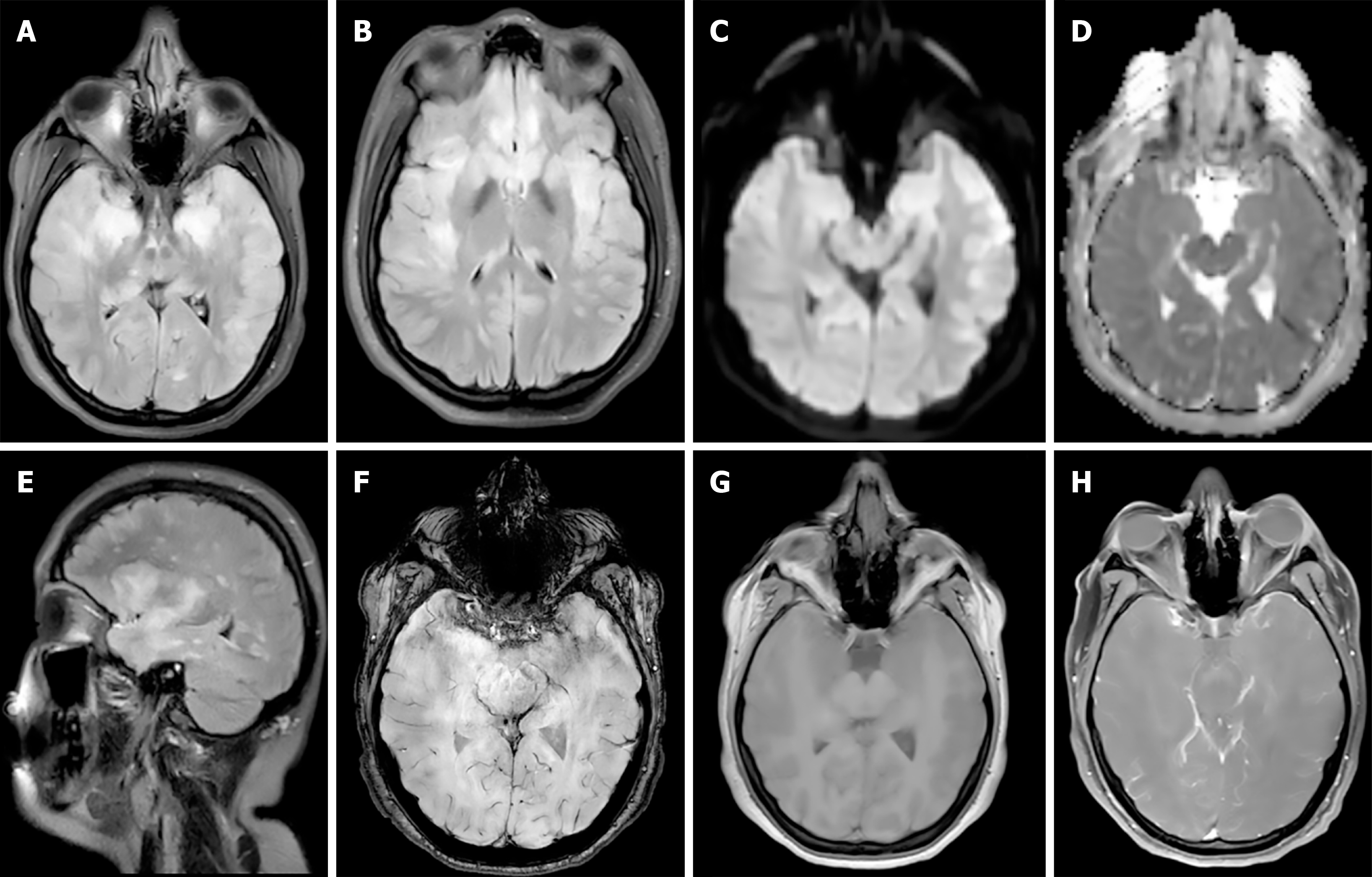
Figure 1 The initial brain magnetic resonance images when the patient was enrolled.
A and B: Axial fluid attenuated inversion recovery imaging; C: Axial diffusion weighted imaging; D: Axial apparent diffusion coefficient imaging; E: Sagittal fluid attenuated inversion recovery imaging; F: Axial susceptibility weighted imaging; G: Axial T1-weighted imaging, pre-contrast; H: Gadolinium-enhanced axial T1-weighted imaging.
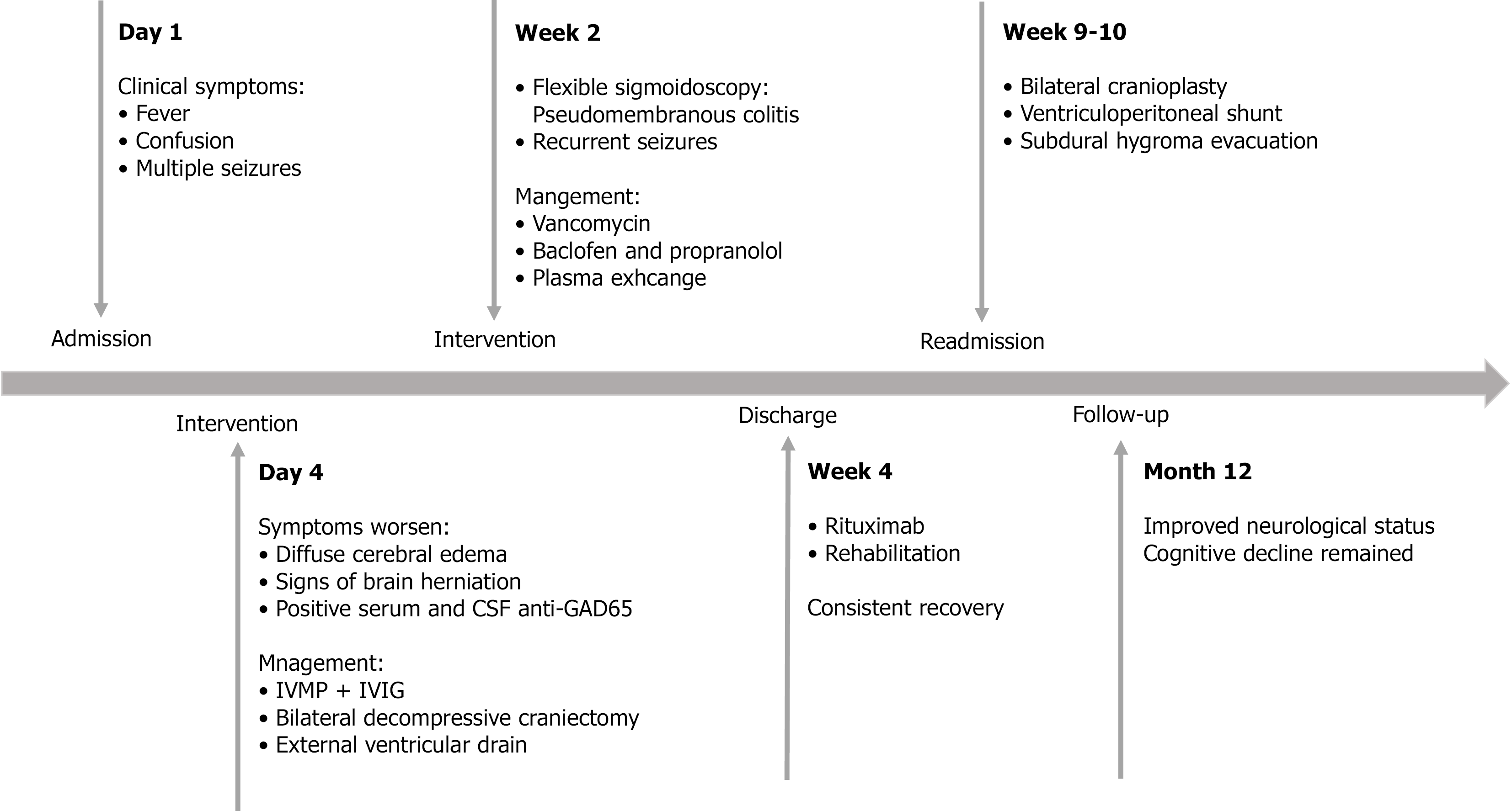
Figure 2 Timeline of symptoms and management of the patient.
CSF: Cerebrospinal fluid; IVIG: Intravenous immunoglobulin; IVMP: Intravenous methylprednisolone.
- Citation: Zhu ZY, Jiang ZR, Chang XC, Xu MY, Zhang X, Zhang B, Wang X. Anti-GAD65 antibody-associated autoimmune encephalitis followed by Clostridium difficile colitis: A case report. World J Transl Med 2026; 12(2): 119153
- URL: https://www.wjgnet.com/2220-6132/full/v12/i2/119153.htm
- DOI: https://dx.doi.org/10.5528/wjtm.119153