©The Author(s) 2019.
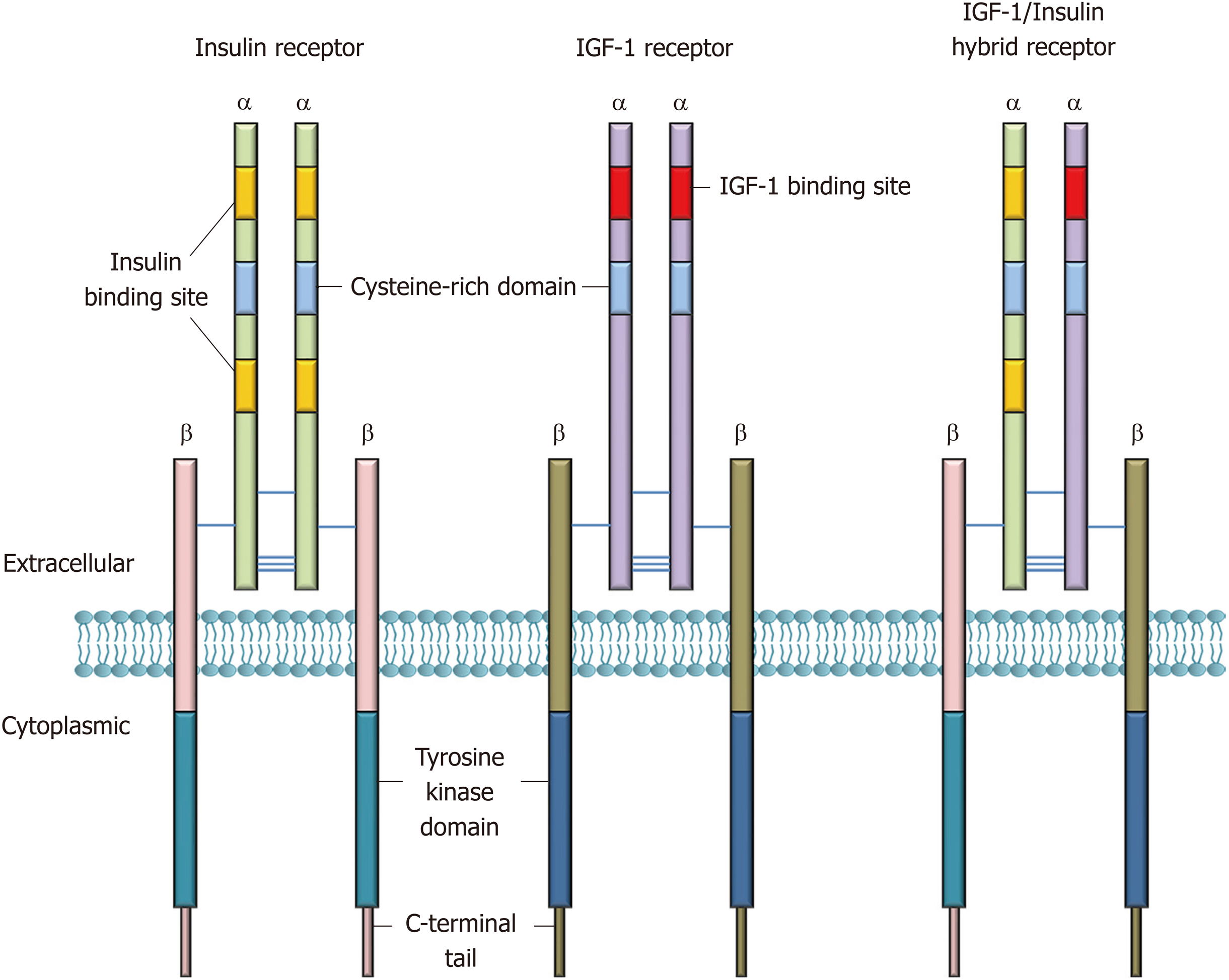
Figure 1 Architecture of insulin and insulin-like growth factor-1 receptors.
Insulin and IGF-1 receptors consist of two extracellular α-chains and two transmembrane β-chains. The α-subunits have binding sites for insulin and IGF-1, whereas the cytoplasmic kinase domain comprises major sites for tyrosine autophosphorylation that are crucial for receptor activation. The α- and β-subunits are connected together via disulfide linkages (Figure is adapted from reference[9]). IGF: Insulin-like growth factor.
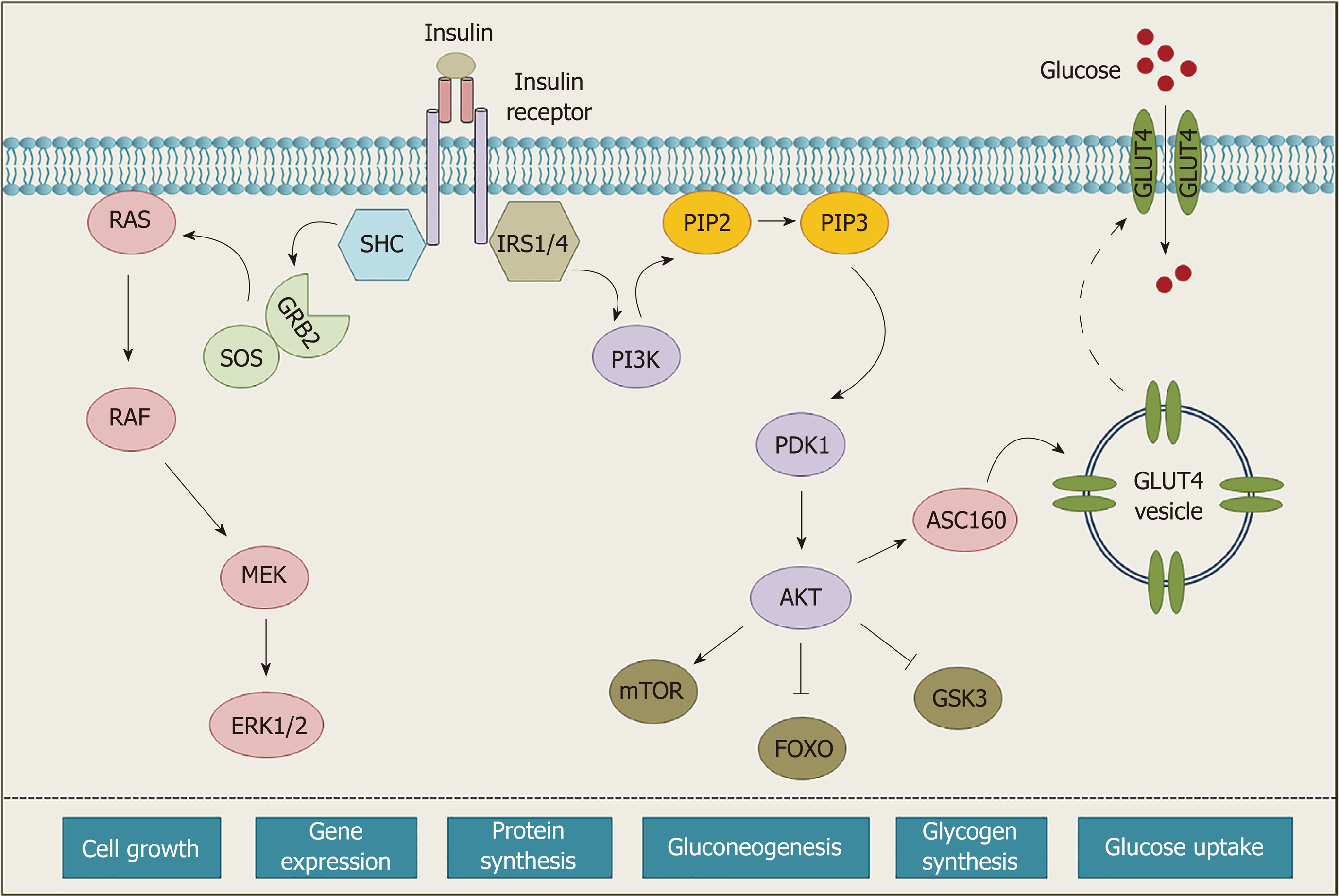
Figure 2 Schematics of the insulin receptor signaling.
Binding of insulin to its receptor causes autophosphorylation of specific tyrosine residues. Upon activation IR recruits different adaptor proteins and initiates a cascade of phosphorylation events. These signaling events ultimately lead to activation or repression of an array of proteins, which regulate various biological functions (Figure is adapted from reference[8]).
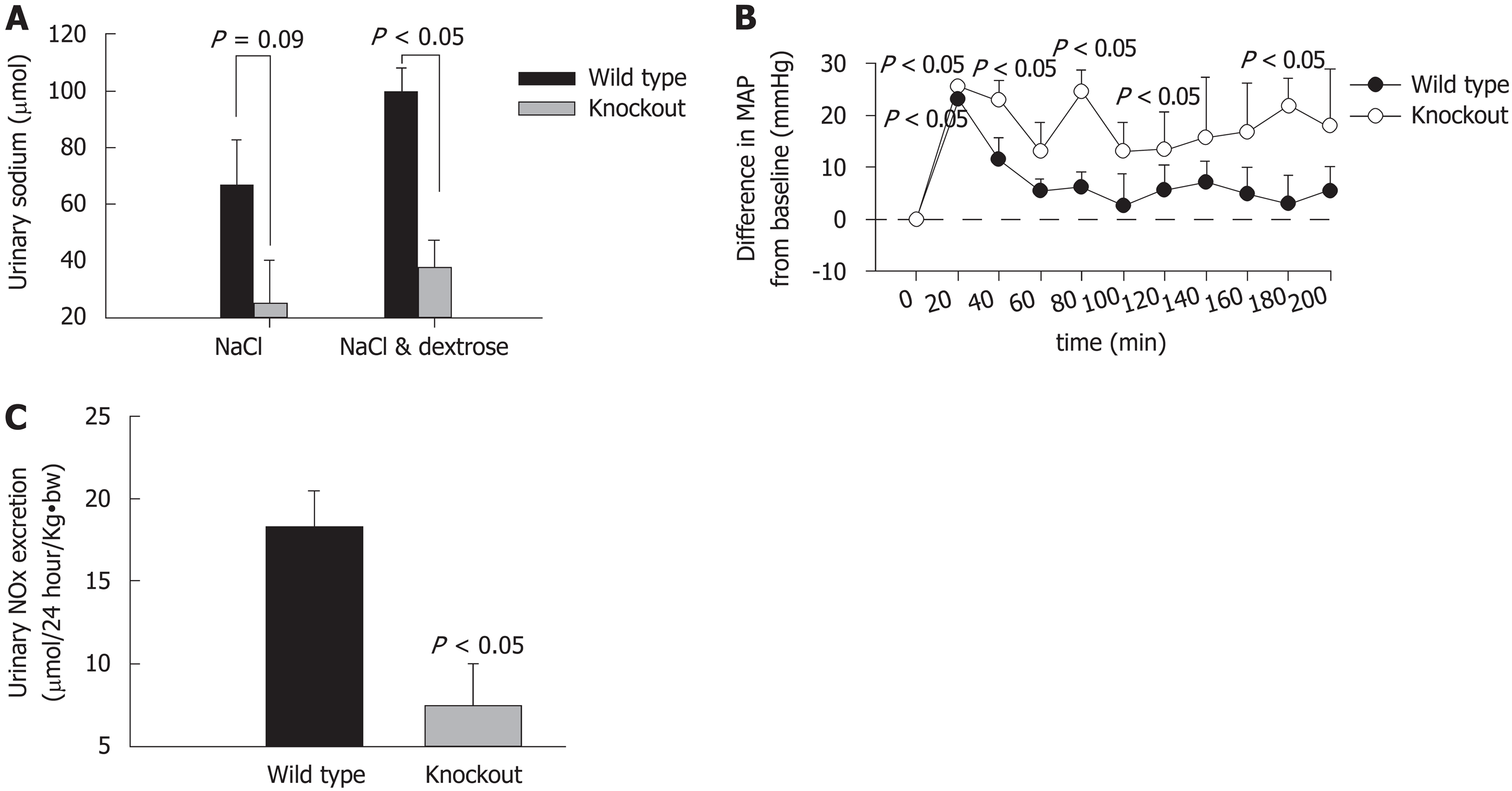
Figure 3 Altered natriuresis and impaired nitric oxide metabolism in insulin receptor-knockout mice.
A: Urinary sodium excretion after oral administration of saline with and without dextrose in 4 h; B: Mean arterial blood pressure (ΔMAP) after NaCl and dextrose administration in mice; C: Urinary nitrate and nitrite excretion in wild-type and insulin receptor-knockout mice after 24 h. (Figure is a modification of figures published in reference[19] and taken with permission).
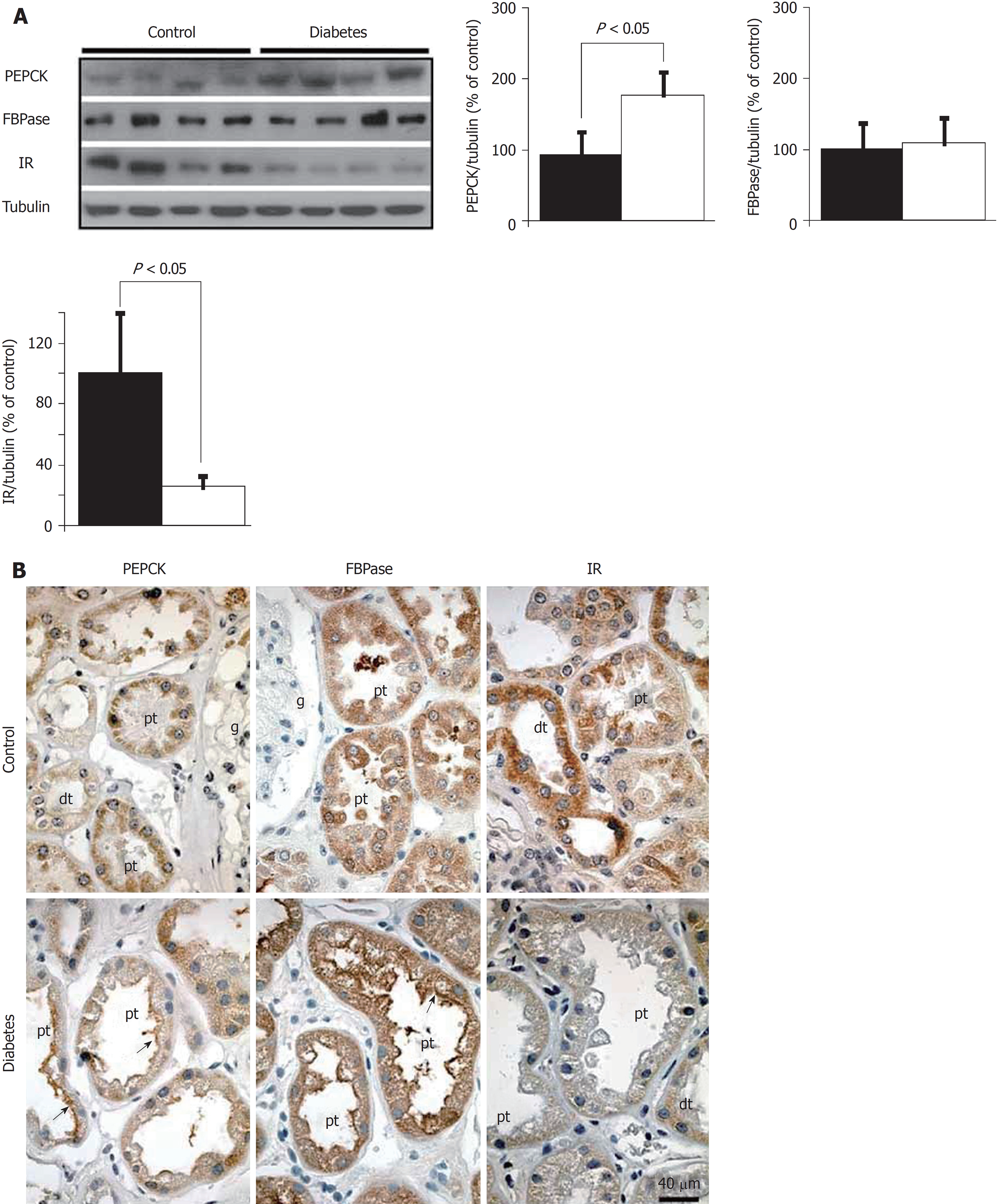
Figure 4 Expression patterns of insulin receptor and gluconeogenic enzymes in normal and diabetic human kidney.
A: Expression of FBPase, PEPCK, IR, and tubulin in renal cortex biopsies of control and Type 2 diabetic individuals analyzed by western blotting; B: Immunohistochemical analysis of FBPase, PEPCK, and IR in renal cortex biopsies of control and Type 2 diabetic individuals (Figure is taken from reference number[6] with permission). PEPCK: Phosphoenolpyruvate carboxykinase; IR: Insulin receptor.
- Citation: Singh S, Sharma R, Kumari M, Tiwari S. Insulin receptors in the kidneys in health and disease. World J Nephrol 2019; 8(1): 11-22
- URL: https://www.wjgnet.com/2220-6124/full/v8/i1/11.htm
- DOI: https://dx.doi.org/10.5527/wjn.v8.i1.11


