©The Author(s) 2015.
World J Nephrol. Nov 6, 2015; 4(5): 480-486
Published online Nov 6, 2015. doi: 10.5527/wjn.v4.i5.480
Published online Nov 6, 2015. doi: 10.5527/wjn.v4.i5.480
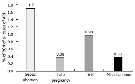
Figure 1 Causes of renal cortical necrosis of all cases of acute renal failure (n = 1822); 1984-2005.
Adapted from Prakash et al[2]. HUS: Hemolytic uremic syndrome; RCN: Renal cortical necrosis.
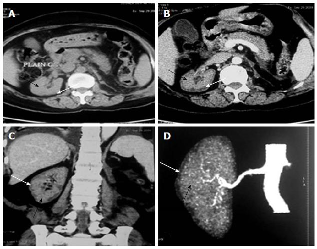
Figure 2 Renal cortical necrosis in a living kidney donor.
Adapted from Prakash et al[32]. Non-contrast (A) computed tomography (CT) scan of abdomen at the level of right hilum showing hypoattenuating peripheral cortical rim (white arrow) as compared to the inner isoattenuating parenchyma (black arrow), On contrast-enhanced axial (B) and coronal (C) scans the central viable parenchyma enhances (black arrow) while the peripheral necrosed cortex does not show any significant enhancement (white arrow). Overall picture is suggesting renal cortical necrosis. The corresponding appearance is also well noted CT Angiogram (D) showing absent uniform nephrogram (white arrow).
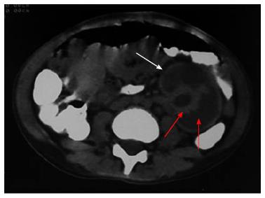
Figure 3 Contrast enhanced computed tomography showing diffuse cortical necrosis in solitary left kidney after road traffic accident in a child aged 11 years.
This image shows isoattenuated peripheral cortical rim (white arrow) and hypoattenuated parenchyma (red arrow).
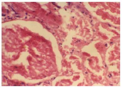
Figure 4 Glomeruli and neighbouring tubules are showing coagulative necrosis.
There is complete loss of nuclei in glomeruli and tubules, but connective tissue framework is preserved. Neutrophilic infiltrations are seen around necrosed glomeruli and in interstitium (HE × 250). Typical histologic feature of renal cortical necrosis.
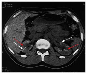
Figure 5 Non contrast computed tomography showing bilateral cortical calcification in a patient with renal cortical necrosis 72 d after acute pancreatitis.
Non contrast computed tomography scan of abdomen at the level of renal hila showing linear hyperattenuation along the renal cortical rim (white arrow) with hypoattenuating medulla (red arrow) in bilateral kidney.
- Citation: Prakash J, Singh VP. Changing picture of renal cortical necrosis in acute kidney injury in developing country. World J Nephrol 2015; 4(5): 480-486
- URL: https://www.wjgnet.com/2220-6124/full/v4/i5/480.htm
- DOI: https://dx.doi.org/10.5527/wjn.v4.i5.480


