©The Author(s) 2015.
World J Nephrol. May 6, 2015; 4(2): 235-244
Published online May 6, 2015. doi: 10.5527/wjn.v4.i2.235
Published online May 6, 2015. doi: 10.5527/wjn.v4.i2.235
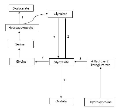
Figure 1 Pathway of oxalate synthesis and enzymatic defects in PH.
A: PH1, alanine glyoxalate aminotransferase; B: PH 2, glycolate reductase hydroxy pyruvate reductase; C: PH 3, 4-hydroxy 2-ketoglutarate aldolase; D: Lactate dehydrogenase.
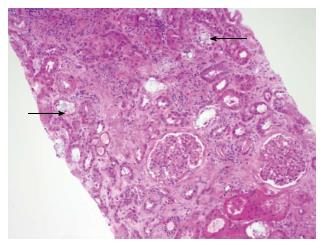
Figure 2 Calcium oxalate deposition in the renal tubules (black arrows).
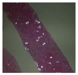
Figure 3 Examination of renal biopsy specimen under polarized light.
Calcium oxalate crystals depict a characteristic birefringence.
- Citation: Bhasin B, Ürekli HM, Atta MG. Primary and secondary hyperoxaluria: Understanding the enigma. World J Nephrol 2015; 4(2): 235-244
- URL: https://www.wjgnet.com/2220-6124/full/v4/i2/235.htm
- DOI: https://dx.doi.org/10.5527/wjn.v4.i2.235


