©The Author(s) 2015.
World J Nephrol. May 6, 2015; 4(2): 169-184
Published online May 6, 2015. doi: 10.5527/wjn.v4.i2.169
Published online May 6, 2015. doi: 10.5527/wjn.v4.i2.169
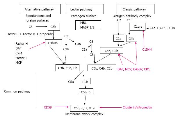
Figure 1 Representation of the classical, lectin and alternative pathways of complement activation, including regulatory molecules (purple).
MBL: Mannose binding lectin; MASP 1/2: Mannan-binding lectin-associated serine protease-1; C1INH: C1 inhibitor; DAF: Decay accelerating factor; CR-1: Complement receptor 1; MCP: Membrane co-factor protein.
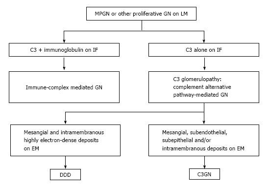
Figure 2 Reclassification of membrano-proliferative glomerulonephritis.
C3GN: C3 glomerulonephritis; DDD: Dense deposit disease; EM: Electron microscopy; GN: Glomerulonephritis; IF: Immunofluorescence; LM: Light microscopy; MPGN: Membrano-proliferative glomerulonephritis.
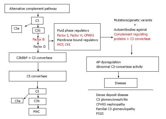
Figure 3 Dysregulation of the alternative complement cascade due to acquired or genetic factors leads to defective complement control causing a range of complement-associated glomerulopathies.
CFHR3: Complement factor H-related protein 3; MCP: Membrane co-factor protein; CR1: Complement receptor 1; AP: Alternative pathway; CFHR5: Complement factor H-related protein 5; FSGS: Focal segments glomerular sclerosis; MAC: Membrane attack complex.
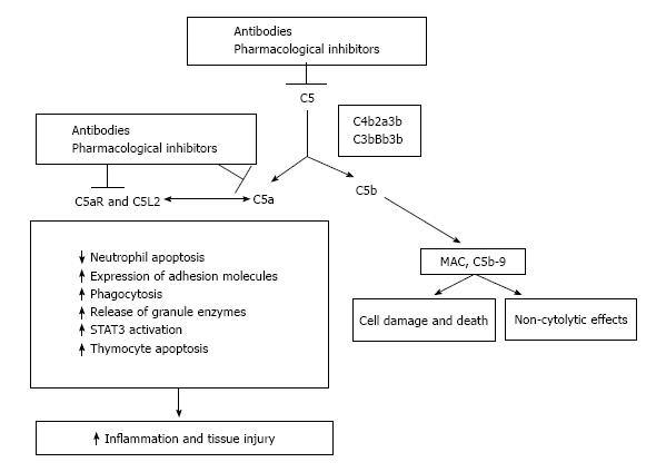
Figure 4 Significance of inhibiting the C5-C5a receptor axis.
C5 convertases formed during activation of complement cascade cleave C5 into its active products. Inhibition is feasible pharmacologically or with neutralizing antibodies. MAC: Membrane attack complex; STAT3: Signal transducers and activators of transcription 3.
- Citation: Salvadori M, Rosso G, Bertoni E. Complement involvement in kidney diseases: From physiopathology to therapeutical targeting. World J Nephrol 2015; 4(2): 169-184
- URL: https://www.wjgnet.com/2220-6124/full/v4/i2/169.htm
- DOI: https://dx.doi.org/10.5527/wjn.v4.i2.169


