
















Copyright: ©Author(s) 2026.
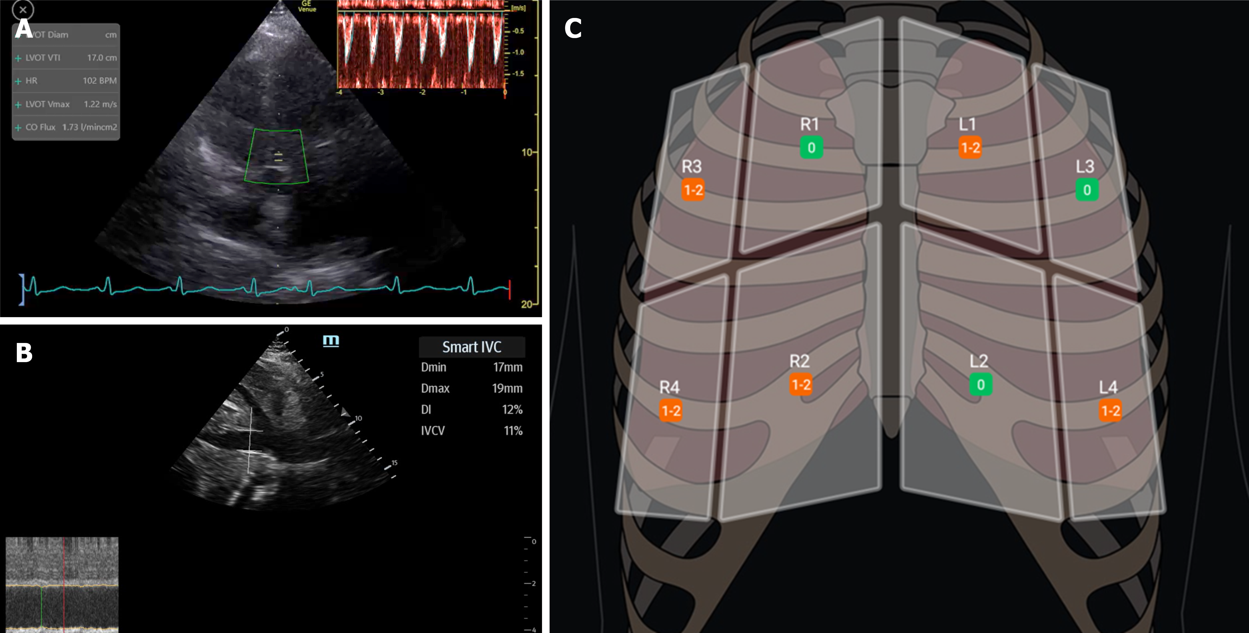
Figure 1 Representative artificial intelligence assisted ultrasound applications in nephrology point-of-care ultrasonography.
A: Automated left ventricular outflow tract velocity-time integral (LVOT VTI) tracing on the GE Venue system (GE HealthCare, Waukesha, WI, United States). Low sweep-speed acquisition as shown here may increase susceptibility to erroneous measurements; B: Smart inferior vena cava (IVC) analysis on the TE7 Max system (Mindray North America, Mahwah, NJ, United States) with automated tracking of longitudinal IVC motion; however, cardiac pulsatility may be incorporated into respiratory variation measurements; C: Automated lung ultrasound B-line summary on the Lumify handheld ultrasound system (Philips Ultrasound, Cambridge, MA, United States). Because each lung zone includes multiple intercostal spaces, serial comparisons may be affected by differences in probe position during follow-up examinations. Dmin: Minimum diameter of the IVC; Dmax: Maximum diameter of the inferior vena cava; DI: Distensibility index; IVCV: Inferior vena cava diameter variation.
- Citation: Koratala A, Ferreira F, Diniz H. Letter to the Editor: Artificial intelligence in nephrology point-of-care ultrasonography - opportunities, limitations, and future directions. World J Nephrol 2026; 15(3): 124607
- URL: https://www.wjgnet.com/2220-6124/full/v15/i3/124607.htm
- DOI: https://dx.doi.org/10.5527/wjn.124607