
















Copyright: ©Author(s) 2026.
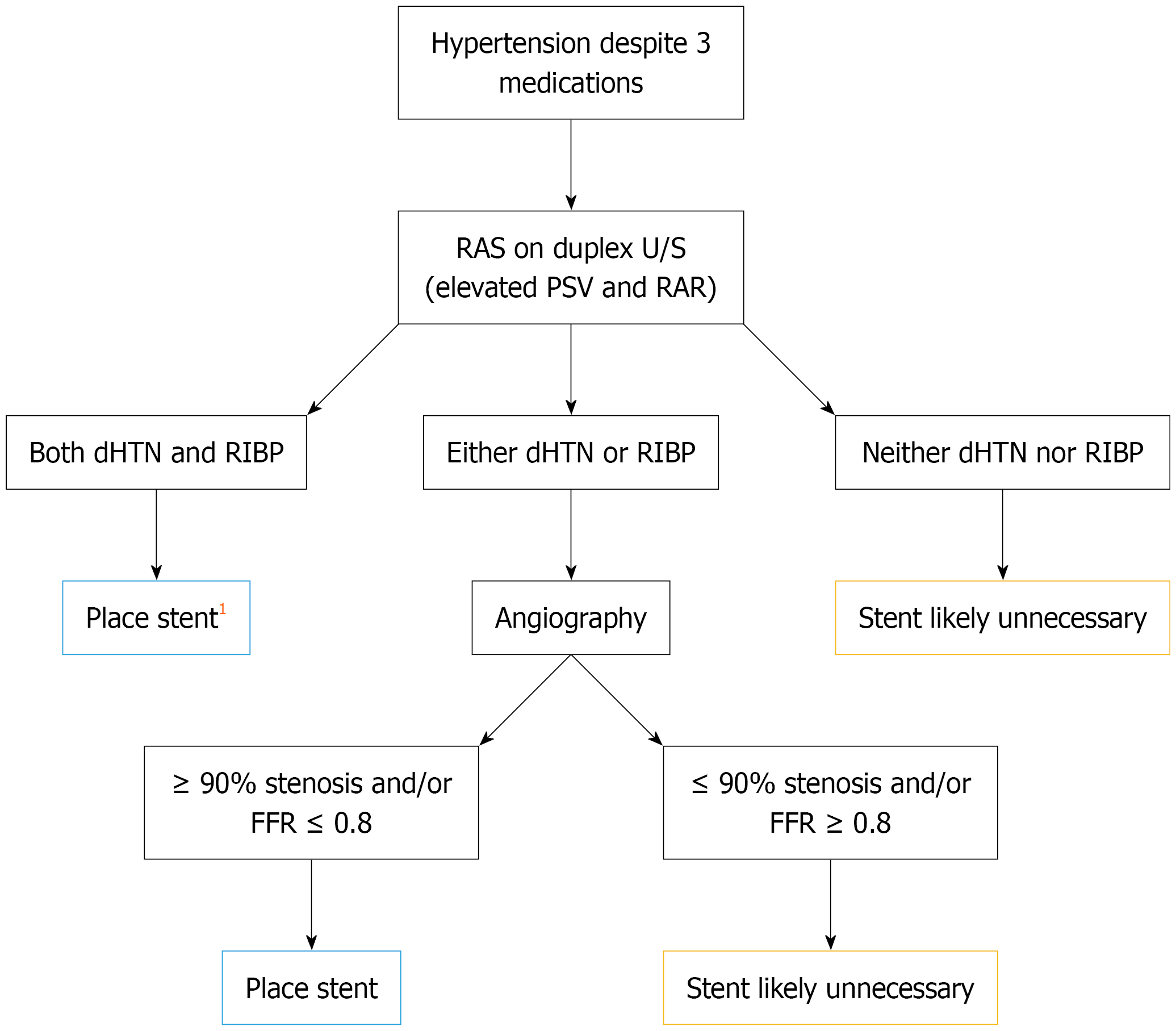
Figure 1 Flow diagram for deciding which patients to stent for resistant hypertension.
When a patient remains hypertensive above 140 mmHg systolic and/or 90 mmHg diastolic despite three medications it is reasonable to get a renal artery duplex ultrasound. If this duplex ultrasound shows renal artery stenosis associated with elevated peak systolic velocity and/or elevated renal-aortic ratio then further history should be evaluated. This should include whether the patient has diastolic hypertension (> 80 mmHg) and/or rapidly increasing blood pressure (systolic blood pressure increase > 15 mmHg within a year). If both are present, then stenting is indicated. 1These patients are highly likely to have ≥ 90% stenosis and/or fractional flow reserve < 0.8. If a patient has either rapidly increasing blood pressure or diastolic hypertension but not both then evaluation for ≥ 90% stenosis and/or fractional flow reserve < 0.8 using angiography is warranted, as is stenting if either are present. If stenosis is ≤ 90% and fractional flow reserve is > 0.8 then stenting is likely unnecessary. If a patient has neither diastolic hypertension or rapidly increasing blood pressure then renal angiography and stenting are likely unnecessary. RAS: Renal artery stenosis; PSV: Peak systolic velocity; RAR: Renal-aortic ratio; dHTN: Diastolic hypertension; RIBP: Rapidly increasing blood pressure; FFR: Fractional flow reserve.
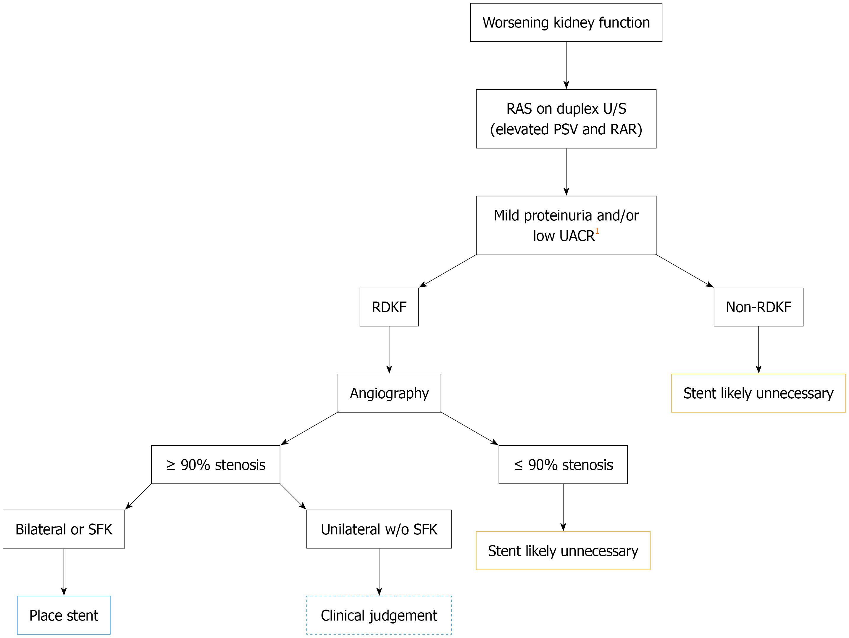
Figure 2 Flow diagram for deciding which patients to stent for ischemic nephropathy.
When a patient is found to have worsening kidney function thought to be related to renal artery disease, the first step is a duplex ultrasound. If this shows elevated peak systolic velocities and renal-aortic ratios, the next step becomes evaluating the health of the kidney parenchyma. This is done by measuring either proteinuria or urine albumin to creatinine ratio. 1It remains unclear what a good cutoff value for these lab tests is but urine albumin to creatinine ratio < 30 mg/g or proteinuria < 1 g per day would indicate healthy parenchyma. Next evaluate for rapidly declining kidney function (RDKF) with glomerular filtration rate trended over the last year. If RDKF is present, then renal angiography is indicated. Without RDKF, stenting is likely unnecessary. If angiography shows a renal artery stenosis ≥ 90% in a patient with a single functioning kidney (congenitally/surgically absent or atrophic contralateral kidney) or in bilateral renal arteries, then stenting is indicated. If the stenosis is ≤ 90% stenting is likely unnecessary. If there is a ≥ 90% stenosis unilaterally in a patient with two functioning kidneys then stenting can be performed but should involve the interventionalists’ clinical judgement about the individual case. RAS: Renal artery stenosis; PSV: Peak systolic velocity; RAR: Renal-aortic ratio; UACR: Urine albumin to creatinine ratio; RDKF: Rapidly declining kidney function; SFK: Single functioning kidney.
- Citation: Warfield AE, Morris CS, Bhave AD, Scriver GM, Massouh A, Kallen J, Solomon RT, Majdalany BS. Renal artery stenting for atherosclerotic renal artery stenosis: A review of contemporary data. World J Nephrol 2026; 15(3): 124524
- URL: https://www.wjgnet.com/2220-6124/full/v15/i3/124524.htm
- DOI: https://dx.doi.org/10.5527/wjn.124524