
















Copyright: ©Author(s) 2026.
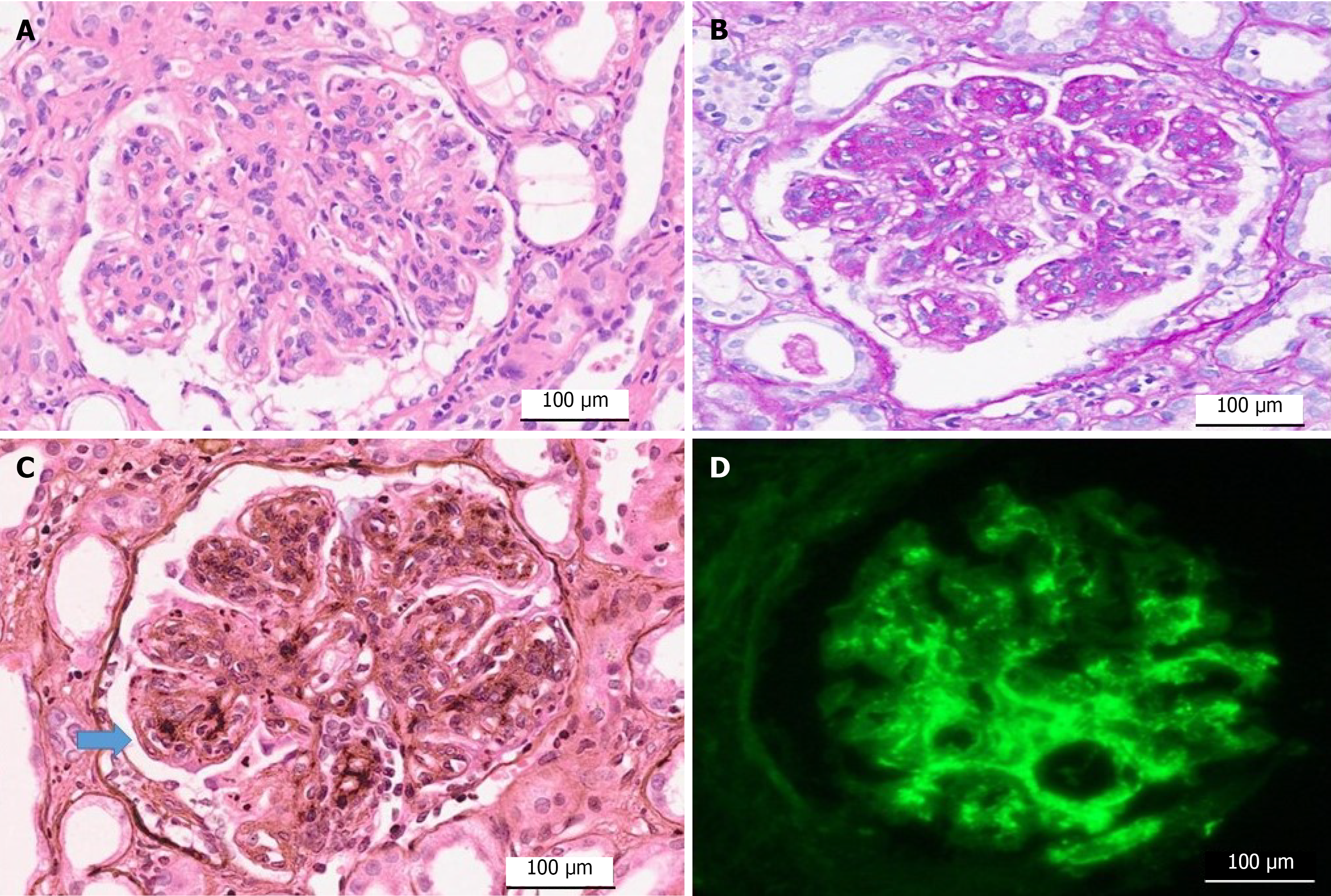
Figure 1 Immunopathology of complement-mediated membranoproliferative glomerulonephritis A: Mesangial and endocapillary hypercellularity with lobular accentuation (hematoxylin and eosin, × 400); B: Mesangial proliferation with almost diffuse inter-positioning into peripheral capillary loops (periodic acid-Schiff stain, × 400); C: Mesangiocapillary proliferation with double contour formation exhibiting tram track appearance (silver methenamine stain, × 400); D: Diffuse, bright, ring-like positivity of C3 with an intensity of 3+ on a scale of 0-3, predominantly in the mesangium on immunofluorescence.
The rest of the immunoreactions were two orders of magnitude lower in intensity (≤ 1+; immunofluorescence, × 400).
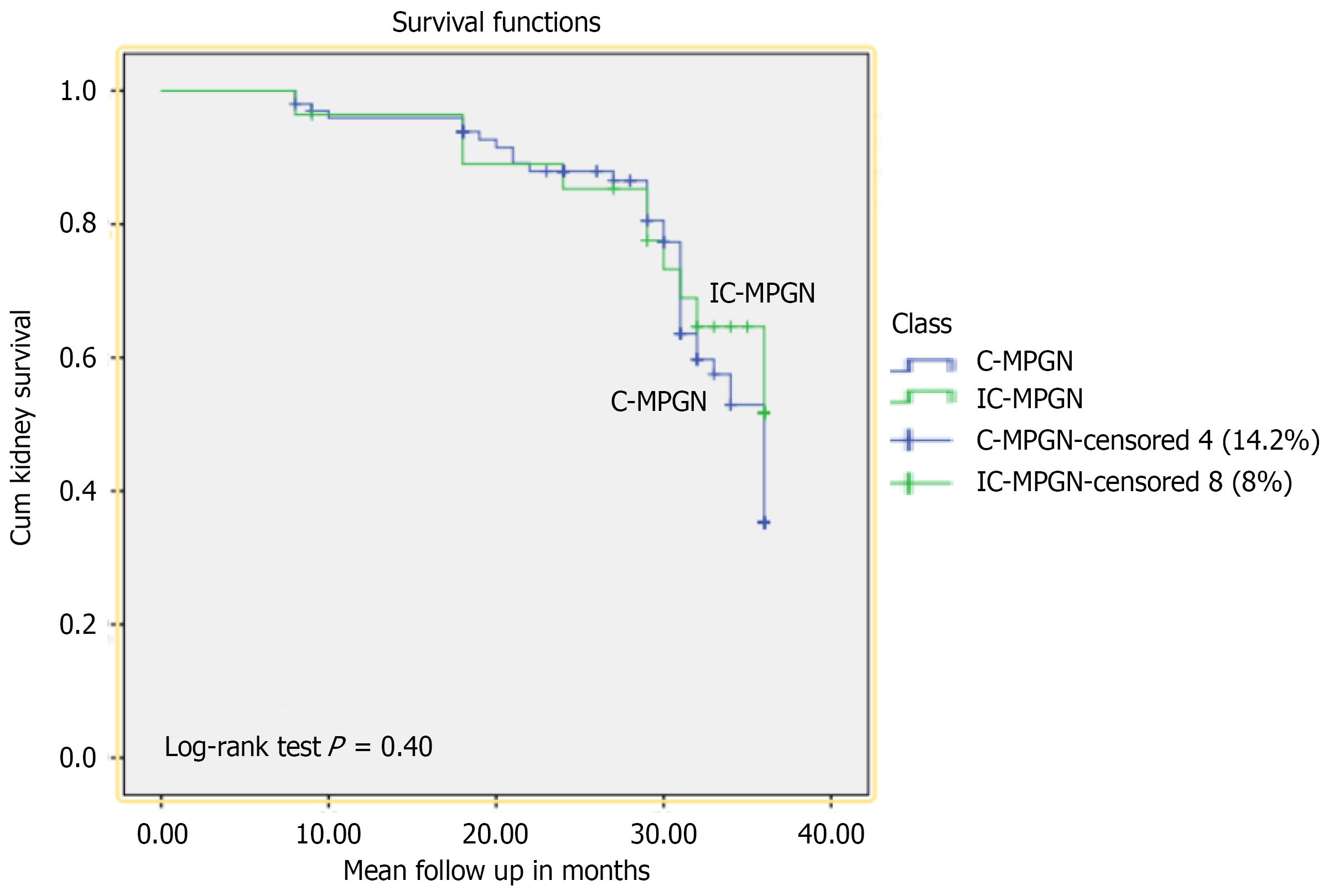
Figure 2 Kaplan-Meier survival analysis curves.
A comparison of survival rates between immune complex-mediated membranoproliferative glome
- Citation: Rashid R, Shrestha RH, Elahi T, Shakeel S, Mubarak M. Immunofluorescence-based reclassification of idiopathic membranoproliferative glomerulonephritis: Insights from a single-center cohort. World J Nephrol 2026; 15(3): 121887
- URL: https://www.wjgnet.com/2220-6124/full/v15/i3/121887.htm
- DOI: https://dx.doi.org/10.5527/wjn.121887